Potassium »
PDB 6f3o-6h6x »
6h6x »
Potassium in PDB 6h6x: Structure of An Evolved Dimeric Form of the Ubid-Class Enzyme Hmff From Pelotomaculum Thermopropionicum in Complex with Prfmn
Protein crystallography data
The structure of Structure of An Evolved Dimeric Form of the Ubid-Class Enzyme Hmff From Pelotomaculum Thermopropionicum in Complex with Prfmn, PDB code: 6h6x
was solved by
K.A.P.Payne,
D.Leys,
with X-Ray Crystallography technique. A brief refinement statistics is given in the table below:
| Resolution Low / High (Å) | 83.86 / 2.25 |
| Space group | P 21 21 21 |
| Cell size a, b, c (Å), α, β, γ (°) | 167.720, 63.930, 98.300, 90.00, 90.00, 90.00 |
| R / Rfree (%) | 19.4 / 24.8 |
Other elements in 6h6x:
The structure of Structure of An Evolved Dimeric Form of the Ubid-Class Enzyme Hmff From Pelotomaculum Thermopropionicum in Complex with Prfmn also contains other interesting chemical elements:
| Manganese | (Mn) | 2 atoms |
| Calcium | (Ca) | 2 atoms |
Potassium Binding Sites:
The binding sites of Potassium atom in the Structure of An Evolved Dimeric Form of the Ubid-Class Enzyme Hmff From Pelotomaculum Thermopropionicum in Complex with Prfmn
(pdb code 6h6x). This binding sites where shown within
5.0 Angstroms radius around Potassium atom.
In total 2 binding sites of Potassium where determined in the Structure of An Evolved Dimeric Form of the Ubid-Class Enzyme Hmff From Pelotomaculum Thermopropionicum in Complex with Prfmn, PDB code: 6h6x:
Jump to Potassium binding site number: 1; 2;
In total 2 binding sites of Potassium where determined in the Structure of An Evolved Dimeric Form of the Ubid-Class Enzyme Hmff From Pelotomaculum Thermopropionicum in Complex with Prfmn, PDB code: 6h6x:
Jump to Potassium binding site number: 1; 2;
Potassium binding site 1 out of 2 in 6h6x
Go back to
Potassium binding site 1 out
of 2 in the Structure of An Evolved Dimeric Form of the Ubid-Class Enzyme Hmff From Pelotomaculum Thermopropionicum in Complex with Prfmn
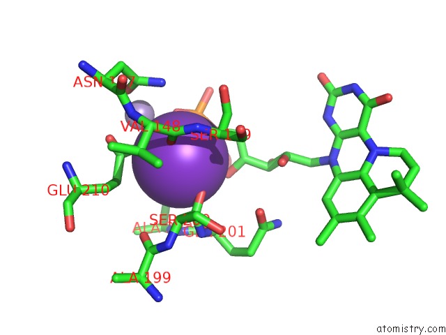
Mono view
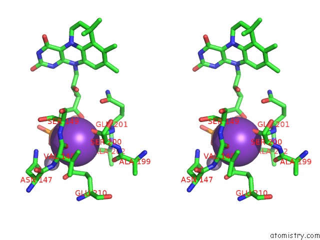
Stereo pair view
Mono view
Stereo pair view
A full contact list of Potassium with other atoms in the K binding
site number 1 of Structure of An Evolved Dimeric Form of the Ubid-Class Enzyme Hmff From Pelotomaculum Thermopropionicum in Complex with Prfmn within 5.0Å range:
|
Potassium binding site 2 out of 2 in 6h6x
Go back to
Potassium binding site 2 out
of 2 in the Structure of An Evolved Dimeric Form of the Ubid-Class Enzyme Hmff From Pelotomaculum Thermopropionicum in Complex with Prfmn
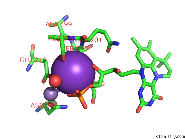
Mono view
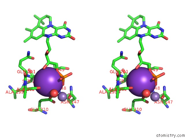
Stereo pair view
Mono view
Stereo pair view
A full contact list of Potassium with other atoms in the K binding
site number 2 of Structure of An Evolved Dimeric Form of the Ubid-Class Enzyme Hmff From Pelotomaculum Thermopropionicum in Complex with Prfmn within 5.0Å range:
|
Reference:
K.A.P.Payne,
S.A.Marshall,
K.Fisher,
M.J.Cliff,
D.M.Cannas,
C.Yan,
D.J.Heyes,
D.A.Parker,
I.Larrosa,
D.Leys.
Enzymatic Carboxylation of 2-Furoic Acid Yields 2,5-Furandicarboxylic Acid (Fdca). Acs Catalysis V. 9 2854 2019.
ISSN: ESSN 2155-5435
PubMed: 31057985
DOI: 10.1021/ACSCATAL.8B04862
Page generated: Mon Aug 12 16:20:55 2024
ISSN: ESSN 2155-5435
PubMed: 31057985
DOI: 10.1021/ACSCATAL.8B04862
Last articles
K in 4LWYK in 4LFC
K in 4LS5
K in 4LP8
K in 4LF6
K in 4LNG
K in 4LGN
K in 4LF8
K in 4LF7
K in 4LFA