Potassium »
PDB 3vuw-3zns »
3wgd »
Potassium in PDB 3wgd: Crystal Structure of ERP46 TRX1
Protein crystallography data
The structure of Crystal Structure of ERP46 TRX1, PDB code: 3wgd
was solved by
K.Inaba,
M.Suzuki,
R.Kojima,
with X-Ray Crystallography technique. A brief refinement statistics is given in the table below:
| Resolution Low / High (Å) | 34.77 / 2.50 |
| Space group | P 1 21 1 |
| Cell size a, b, c (Å), α, β, γ (°) | 50.440, 94.680, 142.020, 90.00, 90.85, 90.00 |
| R / Rfree (%) | 18.8 / 23.5 |
Potassium Binding Sites:
The binding sites of Potassium atom in the Crystal Structure of ERP46 TRX1
(pdb code 3wgd). This binding sites where shown within
5.0 Angstroms radius around Potassium atom.
In total 5 binding sites of Potassium where determined in the Crystal Structure of ERP46 TRX1, PDB code: 3wgd:
Jump to Potassium binding site number: 1; 2; 3; 4; 5;
In total 5 binding sites of Potassium where determined in the Crystal Structure of ERP46 TRX1, PDB code: 3wgd:
Jump to Potassium binding site number: 1; 2; 3; 4; 5;
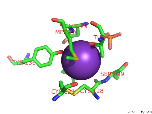
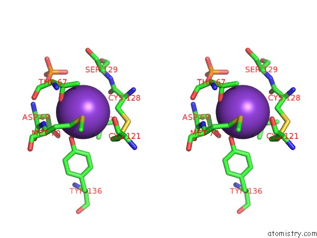
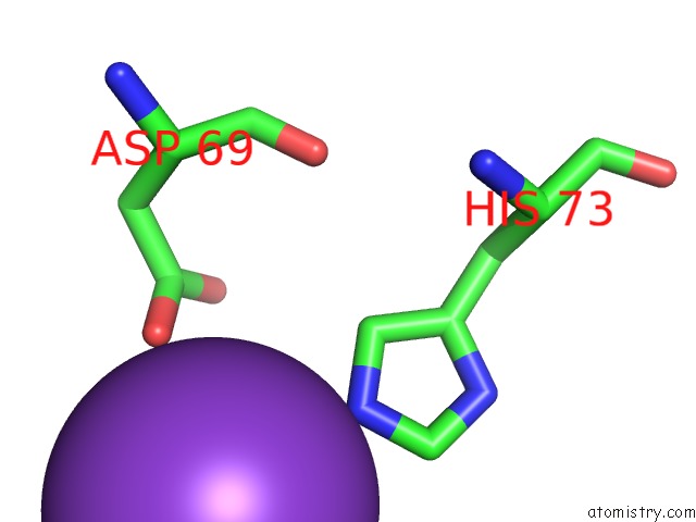
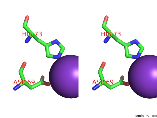
Potassium binding site 1 out of 5 in 3wgd
Go back to
Potassium binding site 1 out
of 5 in the Crystal Structure of ERP46 TRX1
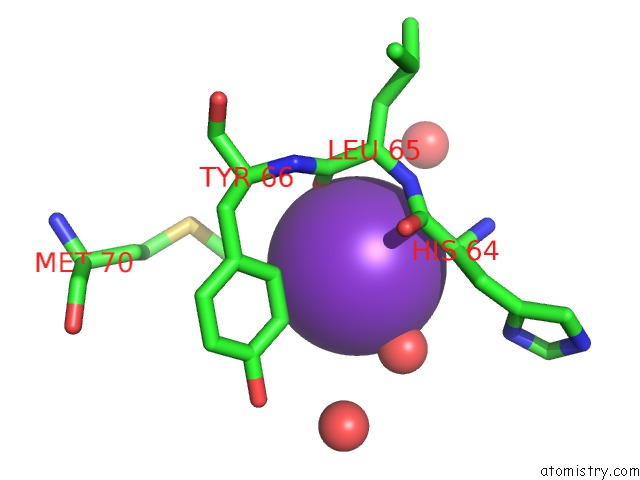
Mono view
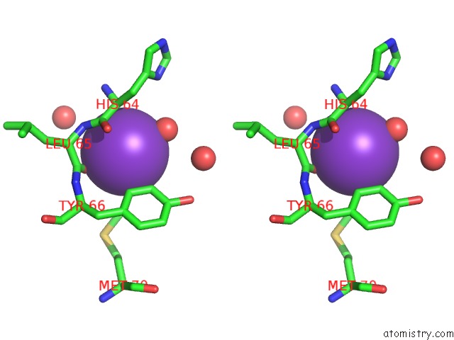
Stereo pair view
Mono view
Stereo pair view
A full contact list of Potassium with other atoms in the K binding
site number 1 of Crystal Structure of ERP46 TRX1 within 5.0Å range:
|
Potassium binding site 2 out of 5 in 3wgd
Go back to
Potassium binding site 2 out
of 5 in the Crystal Structure of ERP46 TRX1
Mono view
Stereo pair view
Mono view
Stereo pair view
A full contact list of Potassium with other atoms in the K binding
site number 2 of Crystal Structure of ERP46 TRX1 within 5.0Å range:
|
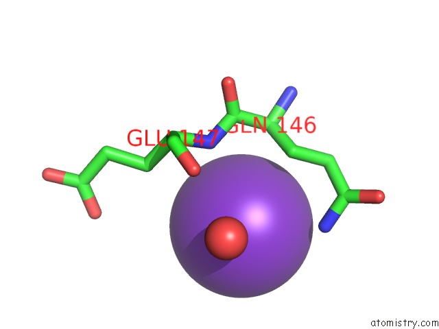
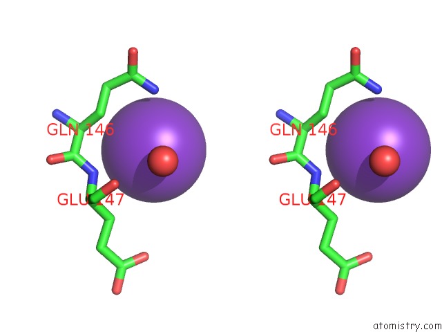
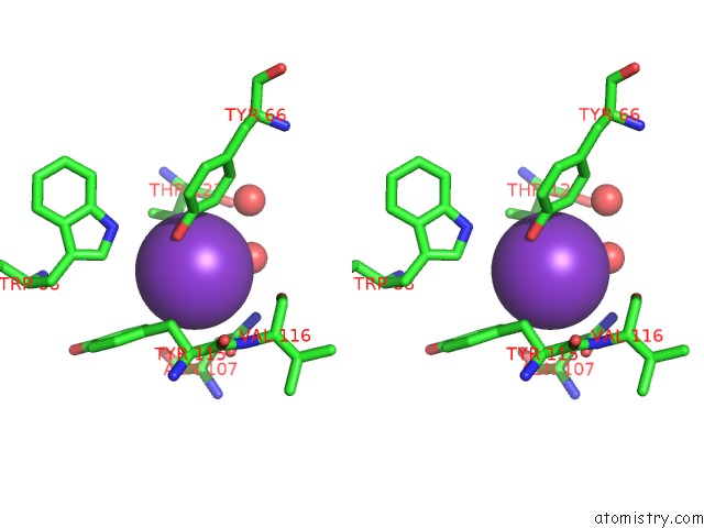
Potassium binding site 3 out of 5 in 3wgd
Go back to
Potassium binding site 3 out
of 5 in the Crystal Structure of ERP46 TRX1
Mono view
Stereo pair view
Mono view
Stereo pair view
A full contact list of Potassium with other atoms in the K binding
site number 3 of Crystal Structure of ERP46 TRX1 within 5.0Å range:
|
Potassium binding site 4 out of 5 in 3wgd
Go back to
Potassium binding site 4 out
of 5 in the Crystal Structure of ERP46 TRX1
Mono view
Stereo pair view
Mono view
Stereo pair view
A full contact list of Potassium with other atoms in the K binding
site number 4 of Crystal Structure of ERP46 TRX1 within 5.0Å range:
|
Potassium binding site 5 out of 5 in 3wgd
Go back to
Potassium binding site 5 out
of 5 in the Crystal Structure of ERP46 TRX1
Mono view
Stereo pair view
Mono view
Stereo pair view
A full contact list of Potassium with other atoms in the K binding
site number 5 of Crystal Structure of ERP46 TRX1 within 5.0Å range:
|
Reference:
R.Kojima,
M.Okumura,
S.Masui,
S.Kanemura,
M.Inoue,
M.Saiki,
H.Yamaguchi,
T.Hikima,
M.Suzuki,
S.Akiyama,
K.Inaba.
Radically Different Thioredoxin Domain Arrangement of ERP46, An Efficient Disulfide Bond Introducer of the Mammalian Pdi Family Structure V. 22 431 2014.
ISSN: ISSN 0969-2126
PubMed: 24462249
DOI: 10.1016/J.STR.2013.12.013
Page generated: Sat Aug 9 06:08:21 2025
ISSN: ISSN 0969-2126
PubMed: 24462249
DOI: 10.1016/J.STR.2013.12.013
Last articles
Na in 2WM2Na in 2WOF
Na in 2WO0
Na in 2WNT
Na in 2WNH
Na in 2WNN
Na in 2WL4
Na in 2WMZ
Na in 2WLU
Na in 2WKV