Potassium »
PDB 3vuw-3zns »
3w8j »
Potassium in PDB 3w8j: Crystal Structure of P5 A0 in A Complex with PRX4 C-Term
Enzymatic activity of Crystal Structure of P5 A0 in A Complex with PRX4 C-Term
All present enzymatic activity of Crystal Structure of P5 A0 in A Complex with PRX4 C-Term:
1.11.1.15; 5.3.4.1;
1.11.1.15; 5.3.4.1;
Protein crystallography data
The structure of Crystal Structure of P5 A0 in A Complex with PRX4 C-Term, PDB code: 3w8j
was solved by
K.Inaba,
M.Suzuki,
R.Kojima,
with X-Ray Crystallography technique. A brief refinement statistics is given in the table below:
| Resolution Low / High (Å) | 41.67 / 2.10 |
| Space group | P 21 21 21 |
| Cell size a, b, c (Å), α, β, γ (°) | 39.045, 53.375, 133.353, 90.00, 90.00, 90.00 |
| R / Rfree (%) | 18.2 / 24.5 |
Potassium Binding Sites:
The binding sites of Potassium atom in the Crystal Structure of P5 A0 in A Complex with PRX4 C-Term
(pdb code 3w8j). This binding sites where shown within
5.0 Angstroms radius around Potassium atom.
In total 4 binding sites of Potassium where determined in the Crystal Structure of P5 A0 in A Complex with PRX4 C-Term, PDB code: 3w8j:
Jump to Potassium binding site number: 1; 2; 3; 4;
In total 4 binding sites of Potassium where determined in the Crystal Structure of P5 A0 in A Complex with PRX4 C-Term, PDB code: 3w8j:
Jump to Potassium binding site number: 1; 2; 3; 4;
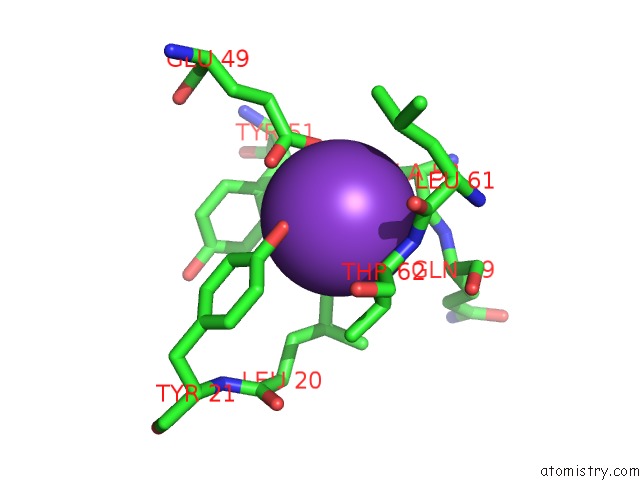
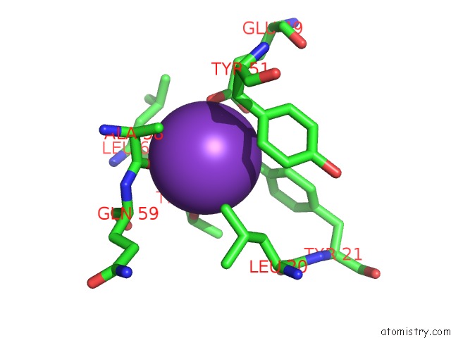
Potassium binding site 1 out of 4 in 3w8j
Go back to
Potassium binding site 1 out
of 4 in the Crystal Structure of P5 A0 in A Complex with PRX4 C-Term
Mono view
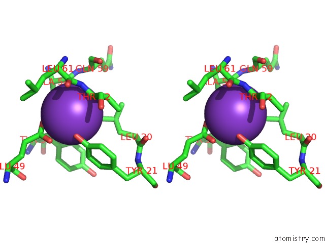
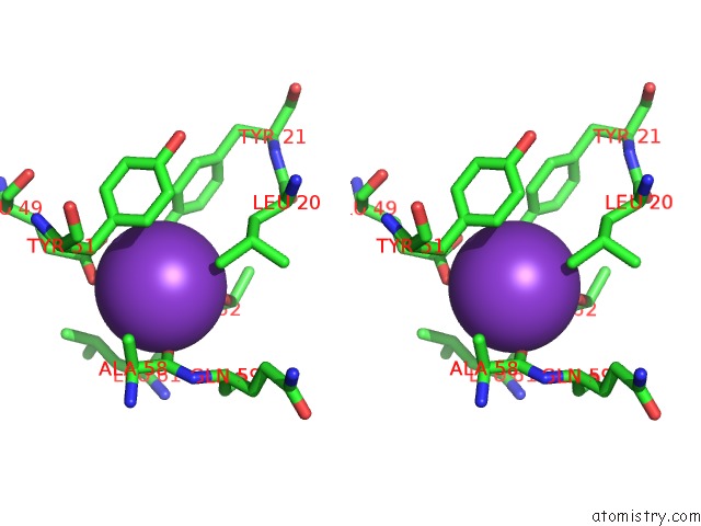
Stereo pair view
Mono view
Stereo pair view
A full contact list of Potassium with other atoms in the K binding
site number 1 of Crystal Structure of P5 A0 in A Complex with PRX4 C-Term within 5.0Å range:
|
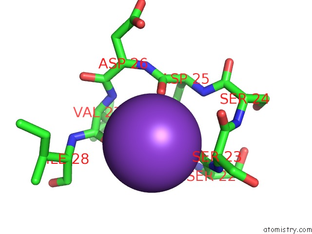
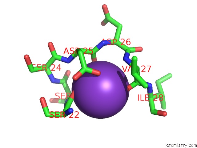
Potassium binding site 2 out of 4 in 3w8j
Go back to
Potassium binding site 2 out
of 4 in the Crystal Structure of P5 A0 in A Complex with PRX4 C-Term
Mono view
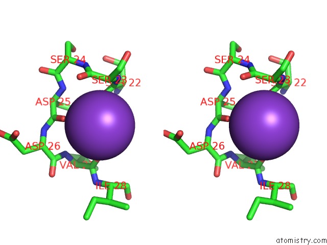
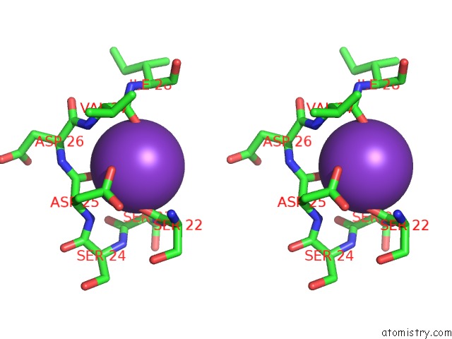
Stereo pair view
Mono view
Stereo pair view
A full contact list of Potassium with other atoms in the K binding
site number 2 of Crystal Structure of P5 A0 in A Complex with PRX4 C-Term within 5.0Å range:
|
Potassium binding site 3 out of 4 in 3w8j
Go back to
Potassium binding site 3 out
of 4 in the Crystal Structure of P5 A0 in A Complex with PRX4 C-Term
Mono view
Stereo pair view
Mono view
Stereo pair view
A full contact list of Potassium with other atoms in the K binding
site number 3 of Crystal Structure of P5 A0 in A Complex with PRX4 C-Term within 5.0Å range:
|
Potassium binding site 4 out of 4 in 3w8j
Go back to
Potassium binding site 4 out
of 4 in the Crystal Structure of P5 A0 in A Complex with PRX4 C-Term
Mono view
Stereo pair view
Mono view
Stereo pair view
A full contact list of Potassium with other atoms in the K binding
site number 4 of Crystal Structure of P5 A0 in A Complex with PRX4 C-Term within 5.0Å range:
|
Reference:
Y.Sato,
R.Kojima,
M.Okumura,
M.Hagiwara,
S.Masui,
K.Maegawa,
M.Saiki,
T.Horibe,
M.Suzuki,
K.Inaba.
Synergistic Cooperation of Pdi Family Members in Peroxiredoxin 4-Driven Oxidative Protein Folding Sci Rep V. 3 2456 2013.
ISSN: ESSN 2045-2322
PubMed: 23949117
DOI: 10.1038/SREP02456
Page generated: Sat Aug 9 06:07:06 2025
ISSN: ESSN 2045-2322
PubMed: 23949117
DOI: 10.1038/SREP02456
Last articles
Mn in 4WZQMn in 4WZM
Mn in 4WYL
Mn in 4WUO
Mn in 4WTM
Mn in 4WTK
Mn in 4WTL
Mn in 4WTJ
Mn in 4WTI
Mn in 4WTF