Potassium »
PDB 1w2b-1yj9 »
1x80 »
Potassium in PDB 1x80: Crystal Structure of the Human Mitochondrial Branched-Chain Alpha- Ketoacid Dehydrogenase
Enzymatic activity of Crystal Structure of the Human Mitochondrial Branched-Chain Alpha- Ketoacid Dehydrogenase
All present enzymatic activity of Crystal Structure of the Human Mitochondrial Branched-Chain Alpha- Ketoacid Dehydrogenase:
1.2.4.4;
1.2.4.4;
Protein crystallography data
The structure of Crystal Structure of the Human Mitochondrial Branched-Chain Alpha- Ketoacid Dehydrogenase, PDB code: 1x80
was solved by
R.M.Wynn,
M.Kato,
M.Machius,
J.L.Chuang,
J.Li,
D.R.Tomchick,
D.T.Chuang,
with X-Ray Crystallography technique. A brief refinement statistics is given in the table below:
| Resolution Low / High (Å) | 50.00 / 2.00 |
| Space group | P 31 2 1 |
| Cell size a, b, c (Å), α, β, γ (°) | 144.403, 144.403, 69.009, 90.00, 90.00, 120.00 |
| R / Rfree (%) | 16 / 19.3 |
Other elements in 1x80:
The structure of Crystal Structure of the Human Mitochondrial Branched-Chain Alpha- Ketoacid Dehydrogenase also contains other interesting chemical elements:
| Manganese | (Mn) | 1 atom |
| Chlorine | (Cl) | 1 atom |
Potassium Binding Sites:
The binding sites of Potassium atom in the Crystal Structure of the Human Mitochondrial Branched-Chain Alpha- Ketoacid Dehydrogenase
(pdb code 1x80). This binding sites where shown within
5.0 Angstroms radius around Potassium atom.
In total 2 binding sites of Potassium where determined in the Crystal Structure of the Human Mitochondrial Branched-Chain Alpha- Ketoacid Dehydrogenase, PDB code: 1x80:
Jump to Potassium binding site number: 1; 2;
In total 2 binding sites of Potassium where determined in the Crystal Structure of the Human Mitochondrial Branched-Chain Alpha- Ketoacid Dehydrogenase, PDB code: 1x80:
Jump to Potassium binding site number: 1; 2;
Potassium binding site 1 out of 2 in 1x80
Go back to
Potassium binding site 1 out
of 2 in the Crystal Structure of the Human Mitochondrial Branched-Chain Alpha- Ketoacid Dehydrogenase
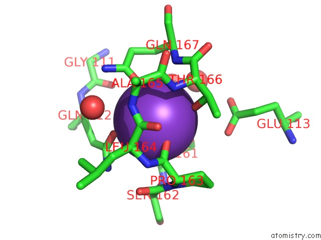
Mono view
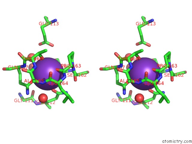
Stereo pair view
Mono view
Stereo pair view
A full contact list of Potassium with other atoms in the K binding
site number 1 of Crystal Structure of the Human Mitochondrial Branched-Chain Alpha- Ketoacid Dehydrogenase within 5.0Å range:
|
Potassium binding site 2 out of 2 in 1x80
Go back to
Potassium binding site 2 out
of 2 in the Crystal Structure of the Human Mitochondrial Branched-Chain Alpha- Ketoacid Dehydrogenase
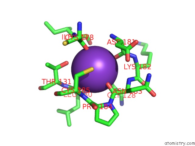
Mono view
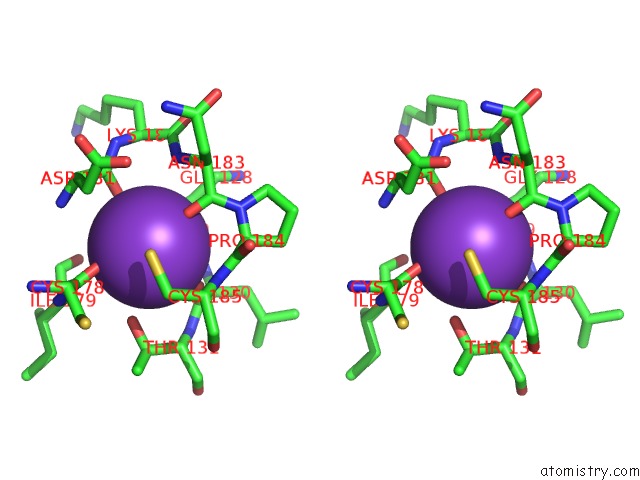
Stereo pair view
Mono view
Stereo pair view
A full contact list of Potassium with other atoms in the K binding
site number 2 of Crystal Structure of the Human Mitochondrial Branched-Chain Alpha- Ketoacid Dehydrogenase within 5.0Å range:
|
Reference:
R.M.Wynn,
M.Kato,
M.Machius,
J.L.Chuang,
J.Li,
D.R.Tomchick,
D.T.Chuang.
Molecular Mechanism For Regulation of the Human Mitochondrial Branched-Chain Alpha-Ketoacid Dehydrogenase Complex By Phosphorylation Structure V. 12 2185 2004.
ISSN: ISSN 0969-2126
PubMed: 15576032
DOI: 10.1016/J.STR.2004.09.013
Page generated: Sat Aug 9 02:54:12 2025
ISSN: ISSN 0969-2126
PubMed: 15576032
DOI: 10.1016/J.STR.2004.09.013
Last articles
K in 3EM2K in 3E8H
K in 3EN2
K in 3EC7
K in 3E8F
K in 3EIY
K in 3E9D
K in 3E9C
K in 3E3S
K in 3E60