Potassium »
PDB 3du0-3f2t »
3ec7 »
Potassium in PDB 3ec7: Crystal Structure of Putative Dehydrogenase From Salmonella Typhimurium LT2
Protein crystallography data
The structure of Crystal Structure of Putative Dehydrogenase From Salmonella Typhimurium LT2, PDB code: 3ec7
was solved by
Y.Kim,
E.Evdokimova,
M.Kudritska,
A.Savchenko,
A.Edwards,
A.Joachimiak,
Midwest Center For Structural Genomics (Mcsg),
with X-Ray Crystallography technique. A brief refinement statistics is given in the table below:
| Resolution Low / High (Å) | 47.00 / 2.15 |
| Space group | P 1 |
| Cell size a, b, c (Å), α, β, γ (°) | 81.001, 98.981, 105.790, 88.05, 81.78, 89.92 |
| R / Rfree (%) | 17.6 / 23.1 |
Potassium Binding Sites:
The binding sites of Potassium atom in the Crystal Structure of Putative Dehydrogenase From Salmonella Typhimurium LT2
(pdb code 3ec7). This binding sites where shown within
5.0 Angstroms radius around Potassium atom.
In total 2 binding sites of Potassium where determined in the Crystal Structure of Putative Dehydrogenase From Salmonella Typhimurium LT2, PDB code: 3ec7:
Jump to Potassium binding site number: 1; 2;
In total 2 binding sites of Potassium where determined in the Crystal Structure of Putative Dehydrogenase From Salmonella Typhimurium LT2, PDB code: 3ec7:
Jump to Potassium binding site number: 1; 2;
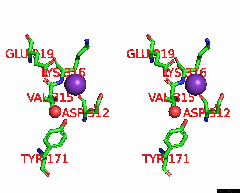
Potassium binding site 1 out of 2 in 3ec7
Go back to
Potassium binding site 1 out
of 2 in the Crystal Structure of Putative Dehydrogenase From Salmonella Typhimurium LT2

Mono view

Stereo pair view
Mono view
Stereo pair view
A full contact list of Potassium with other atoms in the K binding
site number 1 of Crystal Structure of Putative Dehydrogenase From Salmonella Typhimurium LT2 within 5.0Å range:
|
Potassium binding site 2 out of 2 in 3ec7
Go back to
Potassium binding site 2 out
of 2 in the Crystal Structure of Putative Dehydrogenase From Salmonella Typhimurium LT2

Mono view
Stereo pair view
Mono view
Stereo pair view
A full contact list of Potassium with other atoms in the K binding
site number 2 of Crystal Structure of Putative Dehydrogenase From Salmonella Typhimurium LT2 within 5.0Å range:
|
Reference:
Y.Kim,
E.Evdokimova,
M.Kudritska,
A.Savchenko,
A.Edwards,
A.Joachimiak.
Crystal Structure of Putative Dehydrogenase From Salmonella Typhimurium LT2 To Be Published.
Page generated: Sat Aug 9 04:45:02 2025
Last articles
Na in 5ZCKNa in 5ZBI
Na in 5Z96
Na in 5ZB2
Na in 5Z9T
Na in 5Z86
Na in 5Z85
Na in 5Z84
Na in 5Z7W
Na in 5Z48