Potassium »
PDB 7olt-7pji »
7oue »
Potassium in PDB 7oue: Crystal Structure of A Trapped Pab-Agog/Single-Standed Dna Covalent Intermediate
Enzymatic activity of Crystal Structure of A Trapped Pab-Agog/Single-Standed Dna Covalent Intermediate
All present enzymatic activity of Crystal Structure of A Trapped Pab-Agog/Single-Standed Dna Covalent Intermediate:
4.2.99.18;
4.2.99.18;
Protein crystallography data
The structure of Crystal Structure of A Trapped Pab-Agog/Single-Standed Dna Covalent Intermediate, PDB code: 7oue
was solved by
F.Coste,
S.Goffinont,
D.Flament,
B.Castaing,
with X-Ray Crystallography technique. A brief refinement statistics is given in the table below:
| Resolution Low / High (Å) | 71.23 / 2.04 |
| Space group | P 1 |
| Cell size a, b, c (Å), α, β, γ (°) | 39.743, 74.259, 101.726, 92.48, 100.74, 105.46 |
| R / Rfree (%) | 17.5 / 22.3 |
Potassium Binding Sites:
The binding sites of Potassium atom in the Crystal Structure of A Trapped Pab-Agog/Single-Standed Dna Covalent Intermediate
(pdb code 7oue). This binding sites where shown within
5.0 Angstroms radius around Potassium atom.
In total only one binding site of Potassium was determined in the Crystal Structure of A Trapped Pab-Agog/Single-Standed Dna Covalent Intermediate, PDB code: 7oue:
In total only one binding site of Potassium was determined in the Crystal Structure of A Trapped Pab-Agog/Single-Standed Dna Covalent Intermediate, PDB code: 7oue:
Potassium binding site 1 out of 1 in 7oue
Go back to
Potassium binding site 1 out
of 1 in the Crystal Structure of A Trapped Pab-Agog/Single-Standed Dna Covalent Intermediate
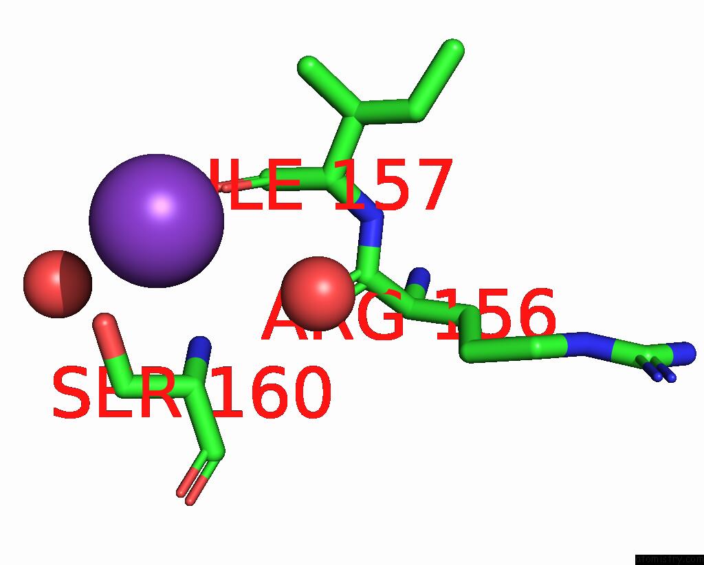
Mono view
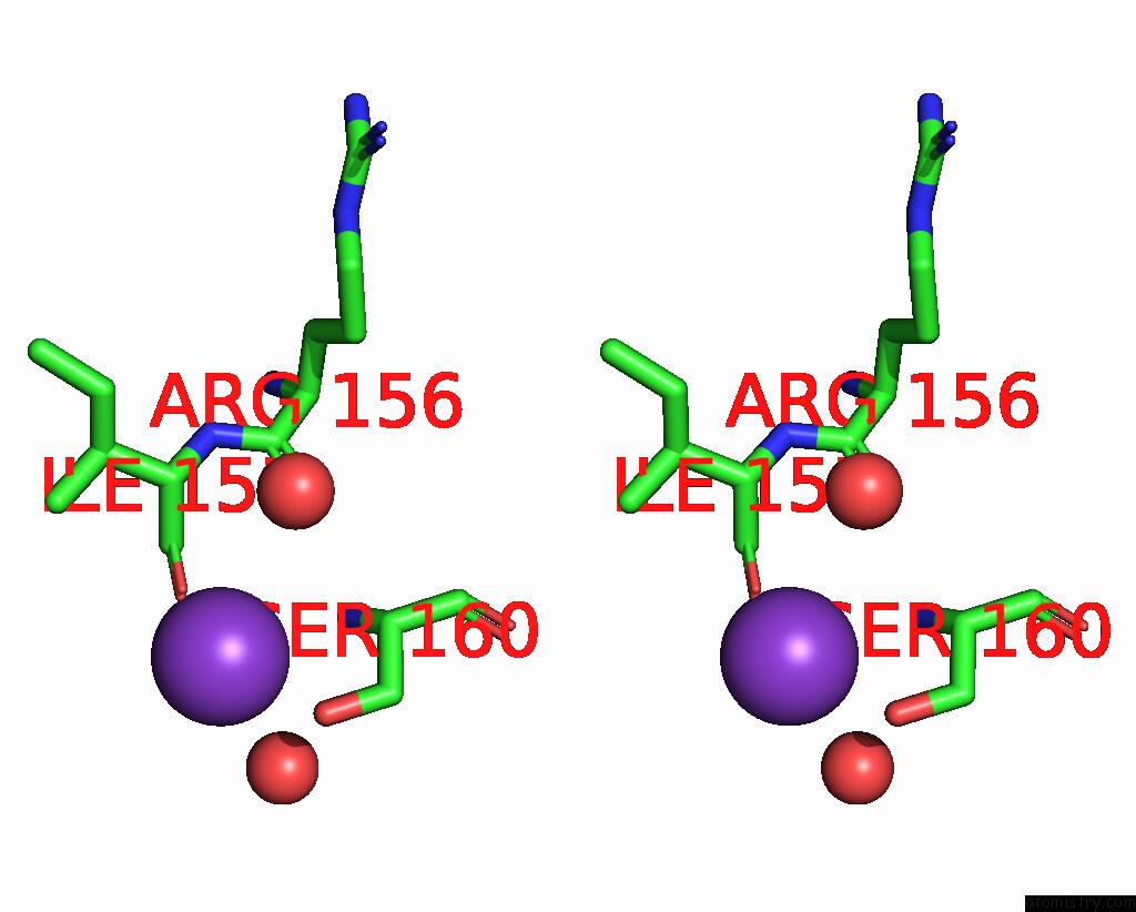
Stereo pair view
Mono view
Stereo pair view
A full contact list of Potassium with other atoms in the K binding
site number 1 of Crystal Structure of A Trapped Pab-Agog/Single-Standed Dna Covalent Intermediate within 5.0Å range:
|
Reference:
F.Coste,
S.Goffinont,
D.Flament,
B.Castaing.
Crystal Structure of A Trapped Pab-Agog/Single-Standed Dna Covalent Intermediate To Be Published.
Page generated: Sat Aug 9 13:53:57 2025
Last articles
K in 8K1EK in 8JZG
K in 8K0U
K in 8K0T
K in 8JGW
K in 8JZA
K in 8JC7
K in 8JGR
K in 8IYM
K in 8JAH