Potassium »
PDB 6n93-6pc3 »
6p9v »
Potassium in PDB 6p9v: Crystal Structure of Hmat Mutant K289L
Enzymatic activity of Crystal Structure of Hmat Mutant K289L
All present enzymatic activity of Crystal Structure of Hmat Mutant K289L:
2.5.1.6;
2.5.1.6;
Protein crystallography data
The structure of Crystal Structure of Hmat Mutant K289L, PDB code: 6p9v
was solved by
M.D.Miller,
W.Xu,
T.D.Huber,
J.A.Clinger,
Y.Liu,
J.S.Thorson,
G.N.Philipsjr.,
with X-Ray Crystallography technique. A brief refinement statistics is given in the table below:
| Resolution Low / High (Å) | 39.74 / 2.05 |
| Space group | I 2 2 2 |
| Cell size a, b, c (Å), α, β, γ (°) | 66.348, 94.624, 116.573, 90.00, 90.00, 90.00 |
| R / Rfree (%) | 17.3 / 20.3 |
Other elements in 6p9v:
The structure of Crystal Structure of Hmat Mutant K289L also contains other interesting chemical elements:
| Magnesium | (Mg) | 1 atom |
Potassium Binding Sites:
The binding sites of Potassium atom in the Crystal Structure of Hmat Mutant K289L
(pdb code 6p9v). This binding sites where shown within
5.0 Angstroms radius around Potassium atom.
In total only one binding site of Potassium was determined in the Crystal Structure of Hmat Mutant K289L, PDB code: 6p9v:
In total only one binding site of Potassium was determined in the Crystal Structure of Hmat Mutant K289L, PDB code: 6p9v:
Potassium binding site 1 out of 1 in 6p9v
Go back to
Potassium binding site 1 out
of 1 in the Crystal Structure of Hmat Mutant K289L
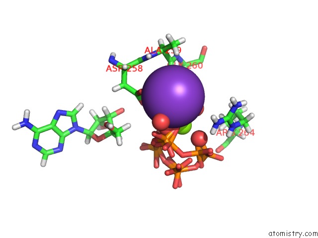
Mono view
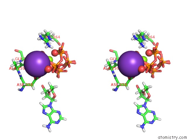
Stereo pair view
Mono view
Stereo pair view
A full contact list of Potassium with other atoms in the K binding
site number 1 of Crystal Structure of Hmat Mutant K289L within 5.0Å range:
|
Reference:
T.D.Huber,
J.A.Clinger,
Y.Liu,
W.Xu,
M.D.Miller,
G.N.Phillips Jr.,
J.S.Thorson.
Methionine Adenosyltransferase Engineering to Enable Bioorthogonal Platforms For Adomet-Utilizing Enzymes. Acs Chem.Biol. V. 15 695 2020.
ISSN: ESSN 1554-8937
PubMed: 32091873
DOI: 10.1021/ACSCHEMBIO.9B00943
Page generated: Sat Aug 9 11:45:26 2025
ISSN: ESSN 1554-8937
PubMed: 32091873
DOI: 10.1021/ACSCHEMBIO.9B00943
Last articles
Mg in 1LNYMg in 1LGH
Mg in 1LNG
Mg in 1L9A
Mg in 1LKX
Mg in 1LM3
Mg in 1LJ0
Mg in 1LJX
Mg in 1LIJ
Mg in 1LII