Potassium in PDB 6p45: Crystal Structure of the G-Quadruplex Formed By (Tgggt)4 in Complex with N-Methylmesoporphryin IX
Protein crystallography data
The structure of Crystal Structure of the G-Quadruplex Formed By (Tgggt)4 in Complex with N-Methylmesoporphryin IX, PDB code: 6p45
was solved by
L.A.Yatsunyk,
L.Y.Lin,
with X-Ray Crystallography technique. A brief refinement statistics is given in the table below:
Resolution Low / High (Å)
|
51.34 /
2.34
|
Space group
|
P 63
|
Cell size a, b, c (Å), α, β, γ (°)
|
59.280,
59.280,
63.332,
90.00,
90.00,
120.00
|
R / Rfree (%)
|
20 /
22.4
|
Potassium Binding Sites:
The binding sites of Potassium atom in the Crystal Structure of the G-Quadruplex Formed By (Tgggt)4 in Complex with N-Methylmesoporphryin IX
(pdb code 6p45). This binding sites where shown within
5.0 Angstroms radius around Potassium atom.
In total 5 binding sites of Potassium where determined in the
Crystal Structure of the G-Quadruplex Formed By (Tgggt)4 in Complex with N-Methylmesoporphryin IX, PDB code: 6p45:
Jump to Potassium binding site number:
1;
2;
3;
4;
5;
Potassium binding site 1 out
of 5 in 6p45
Go back to
Potassium Binding Sites List in 6p45
Potassium binding site 1 out
of 5 in the Crystal Structure of the G-Quadruplex Formed By (Tgggt)4 in Complex with N-Methylmesoporphryin IX
 Mono view
 Stereo pair view
|
A full contact list of Potassium with other atoms in the K binding
site number 1 of Crystal Structure of the G-Quadruplex Formed By (Tgggt)4 in Complex with N-Methylmesoporphryin IX within 5.0Å range:
probe
|
atom
|
residue
|
distance (Å)
|
B
|
Occ
|
A:K101
b:66.9
occ:1.00
|
O6
|
A:DG4
|
2.5
|
66.7
|
1.0
|
O6
|
A:DG8
|
2.7
|
63.3
|
1.0
|
O6
|
A:DG9
|
2.8
|
61.1
|
1.0
|
O6
|
A:DG3
|
2.8
|
57.4
|
1.0
|
O6
|
A:DG13
|
2.8
|
62.6
|
1.0
|
O6
|
A:DG19
|
2.9
|
65.5
|
1.0
|
O6
|
A:DG14
|
2.9
|
65.4
|
1.0
|
O6
|
A:DG18
|
3.0
|
77.9
|
1.0
|
K
|
A:K102
|
3.2
|
59.2
|
1.0
|
C6
|
A:DG4
|
3.5
|
72.4
|
1.0
|
C6
|
A:DG8
|
3.5
|
65.0
|
1.0
|
C6
|
A:DG3
|
3.5
|
68.0
|
1.0
|
C6
|
A:DG13
|
3.7
|
60.5
|
1.0
|
N1
|
A:DG3
|
3.7
|
76.1
|
1.0
|
C6
|
A:DG18
|
3.7
|
73.9
|
1.0
|
C6
|
A:DG19
|
3.7
|
79.8
|
1.0
|
N1
|
A:DG4
|
3.8
|
75.2
|
1.0
|
C6
|
A:DG9
|
3.8
|
64.3
|
1.0
|
N1
|
A:DG8
|
3.8
|
58.9
|
1.0
|
N1
|
A:DG18
|
3.8
|
73.5
|
1.0
|
C6
|
A:DG14
|
3.8
|
75.2
|
1.0
|
N1
|
A:DG19
|
3.9
|
73.4
|
1.0
|
N1
|
A:DG13
|
4.0
|
63.6
|
1.0
|
N1
|
A:DG14
|
4.1
|
82.1
|
1.0
|
N1
|
A:DG9
|
4.1
|
61.3
|
1.0
|
C1
|
A:MMP104
|
4.2
|
80.3
|
1.0
|
C5
|
A:DG8
|
4.7
|
66.5
|
1.0
|
C5
|
A:DG3
|
4.7
|
62.9
|
1.0
|
C5
|
A:DG4
|
4.8
|
74.8
|
1.0
|
C5
|
A:DG13
|
4.9
|
65.9
|
1.0
|
C5
|
A:DG18
|
4.9
|
76.4
|
1.0
|
C2
|
A:DG3
|
4.9
|
73.5
|
1.0
|
|
Potassium binding site 2 out
of 5 in 6p45
Go back to
Potassium Binding Sites List in 6p45
Potassium binding site 2 out
of 5 in the Crystal Structure of the G-Quadruplex Formed By (Tgggt)4 in Complex with N-Methylmesoporphryin IX
 Mono view
 Stereo pair view
|
A full contact list of Potassium with other atoms in the K binding
site number 2 of Crystal Structure of the G-Quadruplex Formed By (Tgggt)4 in Complex with N-Methylmesoporphryin IX within 5.0Å range:
probe
|
atom
|
residue
|
distance (Å)
|
B
|
Occ
|
A:K102
b:59.2
occ:1.00
|
O6
|
A:DG8
|
2.6
|
63.3
|
1.0
|
O6
|
A:DG18
|
2.7
|
77.9
|
1.0
|
O6
|
A:DG17
|
2.7
|
63.8
|
1.0
|
O6
|
A:DG3
|
2.7
|
57.4
|
1.0
|
O6
|
A:DG13
|
2.8
|
62.6
|
1.0
|
O6
|
A:DG12
|
2.8
|
68.1
|
1.0
|
O6
|
A:DG7
|
2.8
|
64.4
|
1.0
|
O6
|
A:DG2
|
3.0
|
68.6
|
1.0
|
K
|
A:K101
|
3.2
|
66.9
|
1.0
|
K
|
A:K103
|
3.4
|
65.5
|
1.0
|
C6
|
A:DG17
|
3.5
|
65.9
|
1.0
|
C6
|
A:DG8
|
3.6
|
65.0
|
1.0
|
C6
|
A:DG18
|
3.6
|
73.9
|
1.0
|
C6
|
A:DG12
|
3.6
|
56.0
|
1.0
|
C6
|
A:DG3
|
3.6
|
68.0
|
1.0
|
C6
|
A:DG13
|
3.6
|
60.5
|
1.0
|
C6
|
A:DG7
|
3.7
|
61.3
|
1.0
|
C6
|
A:DG2
|
3.7
|
71.0
|
1.0
|
N1
|
A:DG18
|
3.8
|
73.5
|
1.0
|
N1
|
A:DG17
|
3.8
|
58.0
|
1.0
|
N1
|
A:DG12
|
3.8
|
58.3
|
1.0
|
N1
|
A:DG8
|
3.8
|
58.9
|
1.0
|
N1
|
A:DG2
|
3.8
|
65.1
|
1.0
|
N1
|
A:DG13
|
3.9
|
63.6
|
1.0
|
N1
|
A:DG3
|
3.9
|
76.1
|
1.0
|
N1
|
A:DG7
|
4.0
|
55.6
|
1.0
|
C5
|
A:DG17
|
4.8
|
66.3
|
1.0
|
C5
|
A:DG12
|
4.8
|
56.6
|
1.0
|
C5
|
A:DG8
|
4.8
|
66.5
|
1.0
|
C5
|
A:DG18
|
4.9
|
76.4
|
1.0
|
C5
|
A:DG7
|
4.9
|
59.5
|
1.0
|
C5
|
A:DG13
|
4.9
|
65.9
|
1.0
|
C5
|
A:DG3
|
4.9
|
62.9
|
1.0
|
C5
|
A:DG2
|
4.9
|
71.4
|
1.0
|
|
Potassium binding site 3 out
of 5 in 6p45
Go back to
Potassium Binding Sites List in 6p45
Potassium binding site 3 out
of 5 in the Crystal Structure of the G-Quadruplex Formed By (Tgggt)4 in Complex with N-Methylmesoporphryin IX
 Mono view
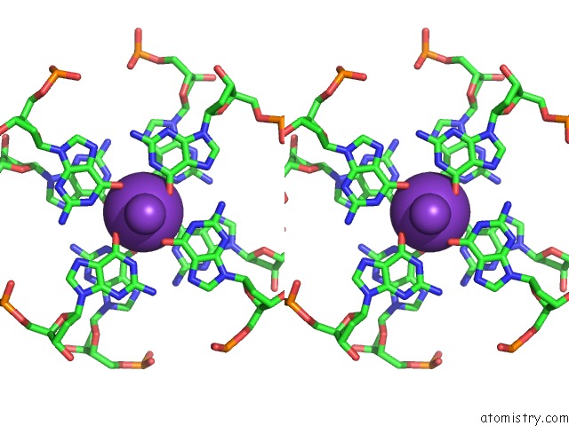
 Stereo pair view
|
A full contact list of Potassium with other atoms in the K binding
site number 3 of Crystal Structure of the G-Quadruplex Formed By (Tgggt)4 in Complex with N-Methylmesoporphryin IX within 5.0Å range:
probe
|
atom
|
residue
|
distance (Å)
|
B
|
Occ
|
A:K103
b:65.5
occ:1.00
|
O6
|
B:DG7
|
2.7
|
56.4
|
1.0
|
O6
|
A:DG12
|
2.7
|
68.1
|
1.0
|
O6
|
A:DG7
|
2.8
|
64.4
|
1.0
|
O6
|
A:DG17
|
2.8
|
63.8
|
1.0
|
O6
|
B:DG2
|
2.8
|
62.8
|
1.0
|
O6
|
B:DG12
|
2.9
|
55.2
|
1.0
|
O6
|
A:DG2
|
2.9
|
68.6
|
1.0
|
O6
|
B:DG17
|
3.0
|
64.9
|
1.0
|
K
|
B:K101
|
3.3
|
58.4
|
1.0
|
K
|
A:K102
|
3.4
|
59.2
|
1.0
|
C6
|
A:DG12
|
3.5
|
56.0
|
1.0
|
C6
|
B:DG7
|
3.5
|
56.8
|
1.0
|
C6
|
A:DG17
|
3.6
|
65.9
|
1.0
|
C6
|
A:DG7
|
3.6
|
61.3
|
1.0
|
C6
|
B:DG2
|
3.7
|
59.0
|
1.0
|
N1
|
A:DG12
|
3.7
|
58.3
|
1.0
|
N1
|
B:DG7
|
3.7
|
50.4
|
1.0
|
C6
|
B:DG17
|
3.7
|
70.3
|
1.0
|
C6
|
B:DG12
|
3.8
|
63.4
|
1.0
|
C6
|
A:DG2
|
3.8
|
71.0
|
1.0
|
N1
|
B:DG2
|
3.8
|
54.7
|
1.0
|
N1
|
B:DG17
|
3.9
|
64.7
|
1.0
|
N1
|
A:DG7
|
3.9
|
55.6
|
1.0
|
N1
|
A:DG17
|
3.9
|
58.0
|
1.0
|
N1
|
A:DG2
|
4.0
|
65.1
|
1.0
|
N1
|
B:DG12
|
4.0
|
68.1
|
1.0
|
C5
|
A:DG12
|
4.7
|
56.6
|
1.0
|
C5
|
B:DG7
|
4.7
|
58.5
|
1.0
|
C5
|
A:DG17
|
4.8
|
66.3
|
1.0
|
C5
|
A:DG7
|
4.9
|
59.5
|
1.0
|
C2
|
A:DG12
|
4.9
|
68.4
|
1.0
|
C5
|
B:DG2
|
5.0
|
68.2
|
1.0
|
C2
|
B:DG7
|
5.0
|
62.7
|
1.0
|
C5
|
B:DG17
|
5.0
|
63.7
|
1.0
|
|
Potassium binding site 4 out
of 5 in 6p45
Go back to
Potassium Binding Sites List in 6p45
Potassium binding site 4 out
of 5 in the Crystal Structure of the G-Quadruplex Formed By (Tgggt)4 in Complex with N-Methylmesoporphryin IX
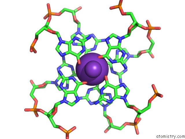
 Mono view
 Stereo pair view
|
A full contact list of Potassium with other atoms in the K binding
site number 4 of Crystal Structure of the G-Quadruplex Formed By (Tgggt)4 in Complex with N-Methylmesoporphryin IX within 5.0Å range:
probe
|
atom
|
residue
|
distance (Å)
|
B
|
Occ
|
B:K101
b:58.4
occ:1.00
|
O6
|
B:DG18
|
2.6
|
80.7
|
1.0
|
O6
|
B:DG3
|
2.7
|
67.0
|
1.0
|
O6
|
B:DG13
|
2.7
|
57.1
|
1.0
|
O6
|
B:DG8
|
2.7
|
57.8
|
1.0
|
O6
|
B:DG7
|
2.8
|
56.4
|
1.0
|
O6
|
B:DG17
|
2.8
|
64.9
|
1.0
|
O6
|
B:DG2
|
2.9
|
62.8
|
1.0
|
O6
|
B:DG12
|
3.0
|
55.2
|
1.0
|
K
|
A:K103
|
3.3
|
65.5
|
1.0
|
K
|
B:K102
|
3.3
|
60.0
|
1.0
|
C6
|
B:DG18
|
3.6
|
67.8
|
1.0
|
C6
|
B:DG7
|
3.6
|
56.8
|
1.0
|
C6
|
B:DG3
|
3.6
|
65.2
|
1.0
|
C6
|
B:DG13
|
3.6
|
63.8
|
1.0
|
C6
|
B:DG17
|
3.6
|
70.3
|
1.0
|
C6
|
B:DG8
|
3.6
|
61.6
|
1.0
|
C6
|
B:DG2
|
3.6
|
59.0
|
1.0
|
N1
|
B:DG2
|
3.8
|
54.7
|
1.0
|
N1
|
B:DG17
|
3.8
|
64.7
|
1.0
|
N1
|
B:DG3
|
3.8
|
60.5
|
1.0
|
C6
|
B:DG12
|
3.8
|
63.4
|
1.0
|
N1
|
B:DG18
|
3.8
|
65.8
|
1.0
|
N1
|
B:DG7
|
3.8
|
50.4
|
1.0
|
N1
|
B:DG8
|
3.8
|
59.8
|
1.0
|
N1
|
B:DG13
|
3.9
|
66.5
|
1.0
|
N1
|
B:DG12
|
4.0
|
68.1
|
1.0
|
C5
|
B:DG7
|
4.8
|
58.5
|
1.0
|
C5
|
B:DG18
|
4.8
|
68.1
|
1.0
|
C5
|
B:DG2
|
4.9
|
68.2
|
1.0
|
C5
|
B:DG3
|
4.9
|
58.1
|
1.0
|
C5
|
B:DG13
|
4.9
|
61.0
|
1.0
|
C5
|
B:DG17
|
4.9
|
63.7
|
1.0
|
C5
|
B:DG8
|
4.9
|
62.9
|
1.0
|
C2
|
B:DG2
|
5.0
|
64.7
|
1.0
|
|
Potassium binding site 5 out
of 5 in 6p45
Go back to
Potassium Binding Sites List in 6p45
Potassium binding site 5 out
of 5 in the Crystal Structure of the G-Quadruplex Formed By (Tgggt)4 in Complex with N-Methylmesoporphryin IX
 Mono view
 Stereo pair view
|
A full contact list of Potassium with other atoms in the K binding
site number 5 of Crystal Structure of the G-Quadruplex Formed By (Tgggt)4 in Complex with N-Methylmesoporphryin IX within 5.0Å range:
probe
|
atom
|
residue
|
distance (Å)
|
B
|
Occ
|
B:K102
b:60.0
occ:1.00
|
O6
|
B:DG4
|
2.7
|
62.9
|
1.0
|
O6
|
B:DG14
|
2.7
|
70.2
|
1.0
|
O6
|
B:DG19
|
2.7
|
60.2
|
1.0
|
O6
|
B:DG9
|
2.8
|
58.6
|
1.0
|
O6
|
B:DG13
|
2.8
|
57.1
|
1.0
|
O6
|
B:DG3
|
2.8
|
67.0
|
1.0
|
O6
|
B:DG8
|
2.9
|
57.8
|
1.0
|
O6
|
B:DG18
|
2.9
|
80.7
|
1.0
|
K
|
B:K101
|
3.3
|
58.4
|
1.0
|
C6
|
B:DG8
|
3.6
|
61.6
|
1.0
|
C6
|
B:DG3
|
3.6
|
65.2
|
1.0
|
C6
|
B:DG18
|
3.6
|
67.8
|
1.0
|
C6
|
B:DG4
|
3.6
|
67.6
|
1.0
|
C6
|
B:DG13
|
3.6
|
63.8
|
1.0
|
C6
|
B:DG19
|
3.7
|
60.6
|
1.0
|
C6
|
B:DG14
|
3.7
|
72.1
|
1.0
|
C6
|
B:DG9
|
3.8
|
60.7
|
1.0
|
N1
|
B:DG8
|
3.8
|
59.8
|
1.0
|
N1
|
B:DG18
|
3.8
|
65.8
|
1.0
|
N1
|
B:DG3
|
3.8
|
60.5
|
1.0
|
N1
|
B:DG19
|
3.9
|
59.9
|
1.0
|
N1
|
B:DG4
|
3.9
|
64.5
|
1.0
|
N1
|
B:DG13
|
4.0
|
66.5
|
1.0
|
N1
|
B:DG14
|
4.0
|
79.7
|
1.0
|
N1
|
B:DG9
|
4.1
|
59.4
|
1.0
|
C5
|
B:DG8
|
4.8
|
62.9
|
1.0
|
N2
|
B:MMP103
|
4.8
|
91.8
|
1.0
|
C5
|
B:DG3
|
4.8
|
58.1
|
1.0
|
C5
|
B:DG18
|
4.8
|
68.1
|
1.0
|
C5
|
B:DG13
|
4.8
|
61.0
|
1.0
|
C5
|
B:DG4
|
4.9
|
66.8
|
1.0
|
C5
|
B:DG19
|
5.0
|
69.1
|
1.0
|
|
Reference:
L.A.Yatsunyk,
L.Y.Lin,
B.Powell.
Biophysical and X-Ray Structural Characterization of the (Gggtt)3GGG G-Quadruplex in Complex with N-Methylmesoporphryin IX To Be Published.
Page generated: Mon Aug 12 17:06:32 2024
|