Potassium »
PDB 6n93-6pc3 »
6ozi »
Potassium in PDB 6ozi: Crystal Structure of Ciona Intestinalis (Ci) Endonuclease V (D234N) in Complex with A 23MER Dna Containing An Inosine Followed By A Ribo- Adenosine
Protein crystallography data
The structure of Crystal Structure of Ciona Intestinalis (Ci) Endonuclease V (D234N) in Complex with A 23MER Dna Containing An Inosine Followed By A Ribo- Adenosine, PDB code: 6ozi
was solved by
N.L.Samara,
W.Yang,
with X-Ray Crystallography technique. A brief refinement statistics is given in the table below:
| Resolution Low / High (Å) | 30.00 / 2.30 |
| Space group | P 2 21 21 |
| Cell size a, b, c (Å), α, β, γ (°) | 51.079, 81.428, 166.034, 90.00, 90.00, 90.00 |
| R / Rfree (%) | 17.4 / 20.1 |
Potassium Binding Sites:
The binding sites of Potassium atom in the Crystal Structure of Ciona Intestinalis (Ci) Endonuclease V (D234N) in Complex with A 23MER Dna Containing An Inosine Followed By A Ribo- Adenosine
(pdb code 6ozi). This binding sites where shown within
5.0 Angstroms radius around Potassium atom.
In total 4 binding sites of Potassium where determined in the Crystal Structure of Ciona Intestinalis (Ci) Endonuclease V (D234N) in Complex with A 23MER Dna Containing An Inosine Followed By A Ribo- Adenosine, PDB code: 6ozi:
Jump to Potassium binding site number: 1; 2; 3; 4;
In total 4 binding sites of Potassium where determined in the Crystal Structure of Ciona Intestinalis (Ci) Endonuclease V (D234N) in Complex with A 23MER Dna Containing An Inosine Followed By A Ribo- Adenosine, PDB code: 6ozi:
Jump to Potassium binding site number: 1; 2; 3; 4;
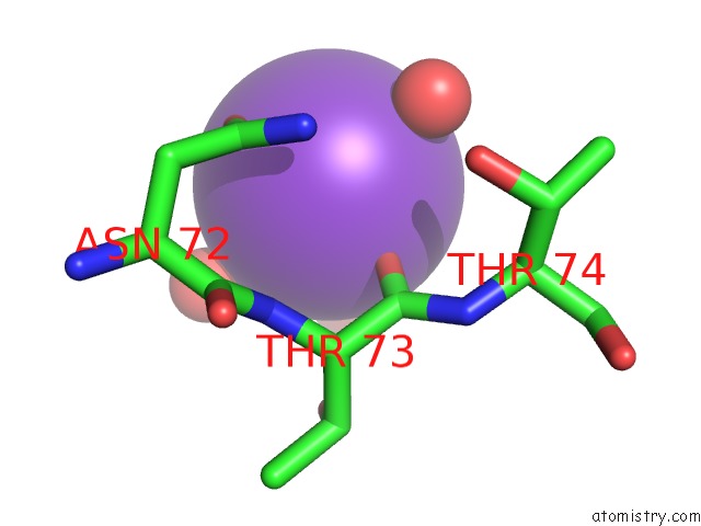
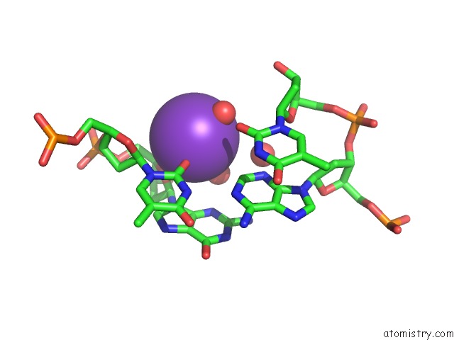
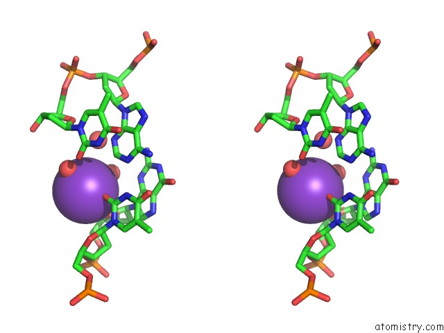
Potassium binding site 1 out of 4 in 6ozi
Go back to
Potassium binding site 1 out
of 4 in the Crystal Structure of Ciona Intestinalis (Ci) Endonuclease V (D234N) in Complex with A 23MER Dna Containing An Inosine Followed By A Ribo- Adenosine
Mono view
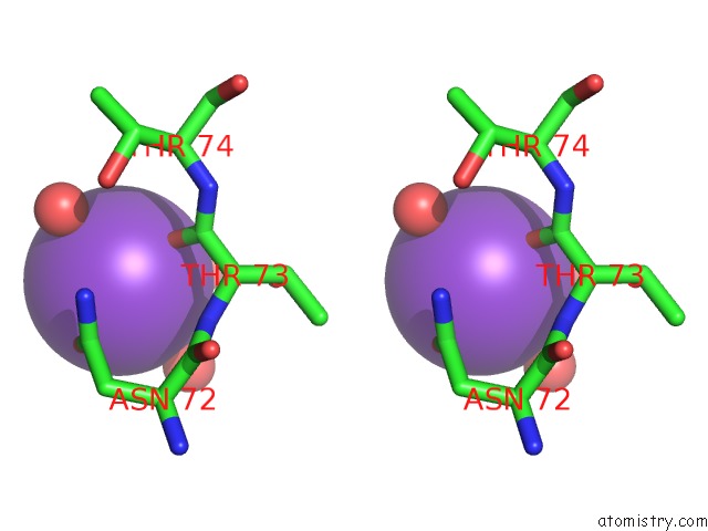
Stereo pair view
Mono view
Stereo pair view
A full contact list of Potassium with other atoms in the K binding
site number 1 of Crystal Structure of Ciona Intestinalis (Ci) Endonuclease V (D234N) in Complex with A 23MER Dna Containing An Inosine Followed By A Ribo- Adenosine within 5.0Å range:
|
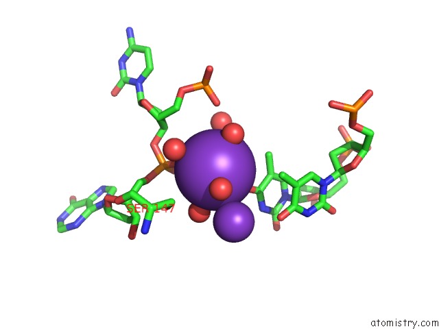
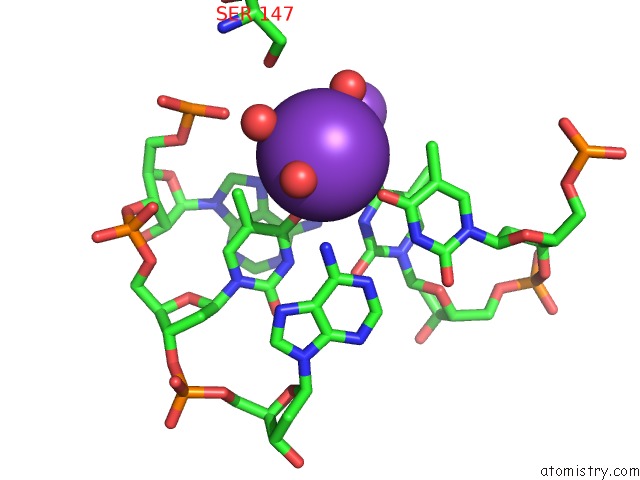
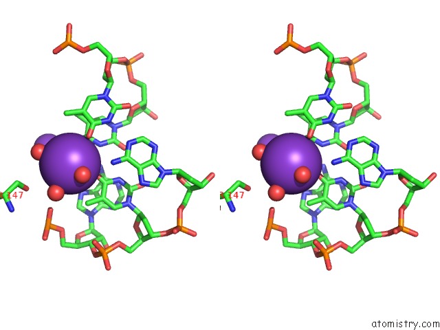
Potassium binding site 2 out of 4 in 6ozi
Go back to
Potassium binding site 2 out
of 4 in the Crystal Structure of Ciona Intestinalis (Ci) Endonuclease V (D234N) in Complex with A 23MER Dna Containing An Inosine Followed By A Ribo- Adenosine
Mono view
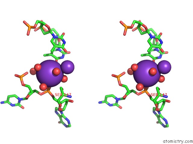
Stereo pair view
Mono view
Stereo pair view
A full contact list of Potassium with other atoms in the K binding
site number 2 of Crystal Structure of Ciona Intestinalis (Ci) Endonuclease V (D234N) in Complex with A 23MER Dna Containing An Inosine Followed By A Ribo- Adenosine within 5.0Å range:
|
Potassium binding site 3 out of 4 in 6ozi
Go back to
Potassium binding site 3 out
of 4 in the Crystal Structure of Ciona Intestinalis (Ci) Endonuclease V (D234N) in Complex with A 23MER Dna Containing An Inosine Followed By A Ribo- Adenosine
Mono view
Stereo pair view
Mono view
Stereo pair view
A full contact list of Potassium with other atoms in the K binding
site number 3 of Crystal Structure of Ciona Intestinalis (Ci) Endonuclease V (D234N) in Complex with A 23MER Dna Containing An Inosine Followed By A Ribo- Adenosine within 5.0Å range:
|
Potassium binding site 4 out of 4 in 6ozi
Go back to
Potassium binding site 4 out
of 4 in the Crystal Structure of Ciona Intestinalis (Ci) Endonuclease V (D234N) in Complex with A 23MER Dna Containing An Inosine Followed By A Ribo- Adenosine
Mono view
Stereo pair view
Mono view
Stereo pair view
A full contact list of Potassium with other atoms in the K binding
site number 4 of Crystal Structure of Ciona Intestinalis (Ci) Endonuclease V (D234N) in Complex with A 23MER Dna Containing An Inosine Followed By A Ribo- Adenosine within 5.0Å range:
|
Reference:
J.Wu,
N.L.Samara,
I.Kuraoka,
W.Yang.
Evolution of Inosine-Specific Endonuclease V From Bacterial Dnase to Eukaryotic Rnase. Mol.Cell V. 76 44 2019.
ISSN: ISSN 1097-2765
PubMed: 31444105
DOI: 10.1016/J.MOLCEL.2019.06.046
Page generated: Mon Aug 12 17:05:33 2024
ISSN: ISSN 1097-2765
PubMed: 31444105
DOI: 10.1016/J.MOLCEL.2019.06.046
Last articles
Zn in 9JYWZn in 9IR4
Zn in 9IR3
Zn in 9GMX
Zn in 9GMW
Zn in 9JEJ
Zn in 9ERF
Zn in 9ERE
Zn in 9EGV
Zn in 9EGW