Potassium in PDB 6c65: Crystal Structure of the Mango-II-A22U Fluorescent Aptamer Bound to TO1-Biotin
Protein crystallography data
The structure of Crystal Structure of the Mango-II-A22U Fluorescent Aptamer Bound to TO1-Biotin, PDB code: 6c65
was solved by
R.J.Trachman,
A.R.Ferre-D'amare,
with X-Ray Crystallography technique. A brief refinement statistics is given in the table below:
Resolution Low / High (Å)
|
46.38 /
2.80
|
Space group
|
C 2 2 21
|
Cell size a, b, c (Å), α, β, γ (°)
|
36.639,
179.680,
108.313,
90.00,
90.00,
90.00
|
R / Rfree (%)
|
18.6 /
23.7
|
Potassium Binding Sites:
The binding sites of Potassium atom in the Crystal Structure of the Mango-II-A22U Fluorescent Aptamer Bound to TO1-Biotin
(pdb code 6c65). This binding sites where shown within
5.0 Angstroms radius around Potassium atom.
In total 8 binding sites of Potassium where determined in the
Crystal Structure of the Mango-II-A22U Fluorescent Aptamer Bound to TO1-Biotin, PDB code: 6c65:
Jump to Potassium binding site number:
1;
2;
3;
4;
5;
6;
7;
8;
Potassium binding site 1 out
of 8 in 6c65
Go back to
Potassium Binding Sites List in 6c65
Potassium binding site 1 out
of 8 in the Crystal Structure of the Mango-II-A22U Fluorescent Aptamer Bound to TO1-Biotin
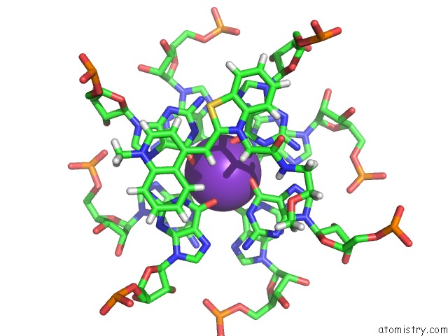
 Mono view
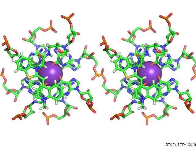
 Stereo pair view
|
A full contact list of Potassium with other atoms in the K binding
site number 1 of Crystal Structure of the Mango-II-A22U Fluorescent Aptamer Bound to TO1-Biotin within 5.0Å range:
probe
|
atom
|
residue
|
distance (Å)
|
B
|
Occ
|
A:K102
b:29.6
occ:1.00
|
O6
|
A:G13
|
2.7
|
35.8
|
1.0
|
O6
|
A:G29
|
2.7
|
37.8
|
1.0
|
O6
|
A:G16
|
2.7
|
35.8
|
1.0
|
O6
|
A:G21
|
2.7
|
38.2
|
1.0
|
O6
|
A:G24
|
2.7
|
44.5
|
1.0
|
O6
|
A:G27
|
2.7
|
35.6
|
1.0
|
O6
|
A:G11
|
2.7
|
33.0
|
1.0
|
O6
|
A:G18
|
2.9
|
42.2
|
1.0
|
K
|
A:K103
|
3.4
|
33.9
|
1.0
|
C6
|
A:G16
|
3.5
|
34.2
|
1.0
|
C6
|
A:G13
|
3.5
|
35.9
|
1.0
|
C6
|
A:G27
|
3.5
|
32.1
|
1.0
|
C6
|
A:G24
|
3.5
|
43.1
|
1.0
|
C6
|
A:G29
|
3.6
|
34.5
|
1.0
|
C6
|
A:G21
|
3.6
|
38.7
|
1.0
|
C6
|
A:G18
|
3.6
|
38.7
|
1.0
|
C6
|
A:G11
|
3.6
|
31.1
|
1.0
|
N1
|
A:G27
|
3.7
|
33.3
|
1.0
|
N1
|
A:G13
|
3.8
|
31.8
|
1.0
|
N1
|
A:G18
|
3.9
|
39.6
|
1.0
|
N1
|
A:G29
|
3.9
|
32.1
|
1.0
|
N1
|
A:G16
|
3.9
|
35.5
|
1.0
|
N1
|
A:G24
|
3.9
|
41.6
|
1.0
|
N1
|
A:G21
|
3.9
|
38.3
|
1.0
|
N1
|
A:G11
|
4.0
|
33.1
|
1.0
|
H082
|
A:EKJ101
|
4.5
|
57.5
|
1.0
|
C5
|
A:G24
|
4.7
|
41.4
|
1.0
|
C5
|
A:G16
|
4.7
|
29.7
|
1.0
|
C5
|
A:G13
|
4.8
|
38.5
|
1.0
|
C5
|
A:G27
|
4.8
|
30.9
|
1.0
|
C5
|
A:G11
|
4.8
|
28.8
|
1.0
|
C5
|
A:G18
|
4.8
|
37.7
|
1.0
|
C5
|
A:G29
|
4.8
|
33.9
|
1.0
|
C5
|
A:G21
|
4.9
|
39.2
|
1.0
|
H111
|
A:EKJ101
|
4.9
|
61.9
|
1.0
|
C2
|
A:G27
|
5.0
|
31.6
|
1.0
|
|
Potassium binding site 2 out
of 8 in 6c65
Go back to
Potassium Binding Sites List in 6c65
Potassium binding site 2 out
of 8 in the Crystal Structure of the Mango-II-A22U Fluorescent Aptamer Bound to TO1-Biotin
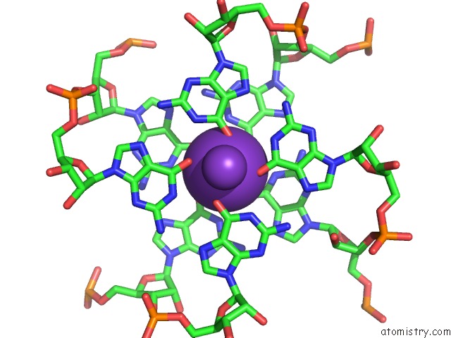
 Mono view
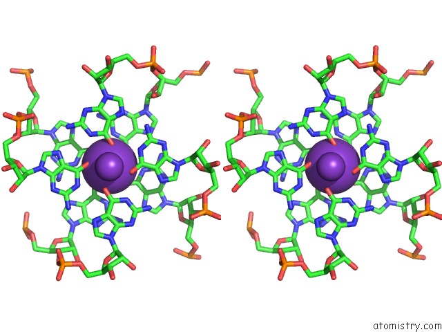
 Stereo pair view
|
A full contact list of Potassium with other atoms in the K binding
site number 2 of Crystal Structure of the Mango-II-A22U Fluorescent Aptamer Bound to TO1-Biotin within 5.0Å range:
probe
|
atom
|
residue
|
distance (Å)
|
B
|
Occ
|
A:K103
b:33.9
occ:1.00
|
O6
|
A:G16
|
2.6
|
35.8
|
1.0
|
O6
|
A:G20
|
2.7
|
33.5
|
1.0
|
O6
|
A:G26
|
2.7
|
39.5
|
1.0
|
O6
|
A:G11
|
2.7
|
33.0
|
1.0
|
O6
|
A:G15
|
2.8
|
35.4
|
1.0
|
O6
|
A:G27
|
2.8
|
35.6
|
1.0
|
O6
|
A:G10
|
2.9
|
30.8
|
1.0
|
O6
|
A:G21
|
3.0
|
38.2
|
1.0
|
K
|
A:K104
|
3.3
|
47.7
|
0.5
|
K
|
A:K102
|
3.4
|
29.6
|
1.0
|
C6
|
A:G10
|
3.5
|
30.9
|
1.0
|
C6
|
A:G16
|
3.6
|
34.2
|
1.0
|
C6
|
A:G15
|
3.6
|
31.5
|
1.0
|
C6
|
A:G27
|
3.6
|
32.1
|
1.0
|
C6
|
A:G26
|
3.6
|
33.2
|
1.0
|
C6
|
A:G20
|
3.6
|
36.8
|
1.0
|
C6
|
A:G11
|
3.6
|
31.1
|
1.0
|
N1
|
A:G15
|
3.7
|
28.4
|
1.0
|
N1
|
A:G10
|
3.7
|
28.2
|
1.0
|
N1
|
A:G26
|
3.8
|
33.4
|
1.0
|
N1
|
A:G27
|
3.8
|
33.3
|
1.0
|
C6
|
A:G21
|
3.8
|
38.7
|
1.0
|
N1
|
A:G20
|
3.9
|
38.9
|
1.0
|
N1
|
A:G11
|
3.9
|
33.1
|
1.0
|
N1
|
A:G16
|
4.0
|
35.5
|
1.0
|
N1
|
A:G21
|
4.0
|
38.3
|
1.0
|
C5
|
A:G10
|
4.7
|
30.6
|
1.0
|
C5
|
A:G16
|
4.7
|
29.7
|
1.0
|
C5
|
A:G27
|
4.7
|
30.9
|
1.0
|
C2
|
A:G10
|
4.8
|
30.8
|
1.0
|
C5
|
A:G26
|
4.8
|
33.1
|
1.0
|
C5
|
A:G15
|
4.8
|
31.4
|
1.0
|
C5
|
A:G11
|
4.9
|
28.8
|
1.0
|
C5
|
A:G20
|
4.9
|
37.0
|
1.0
|
C2
|
A:G15
|
5.0
|
29.7
|
1.0
|
|
Potassium binding site 3 out
of 8 in 6c65
Go back to
Potassium Binding Sites List in 6c65
Potassium binding site 3 out
of 8 in the Crystal Structure of the Mango-II-A22U Fluorescent Aptamer Bound to TO1-Biotin
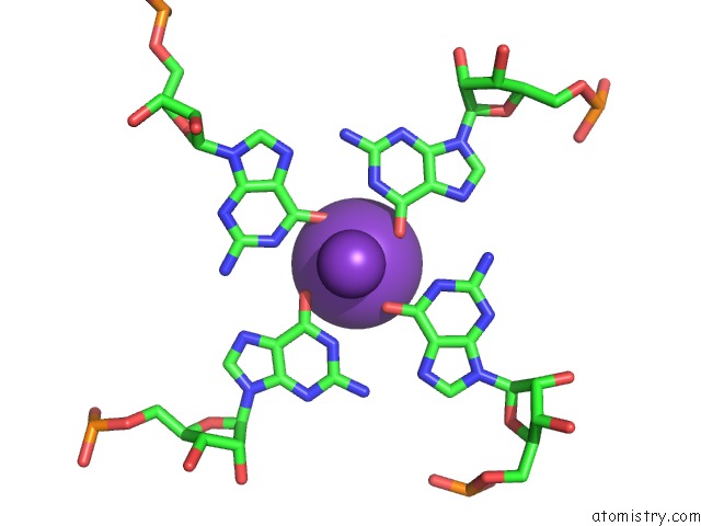
 Mono view
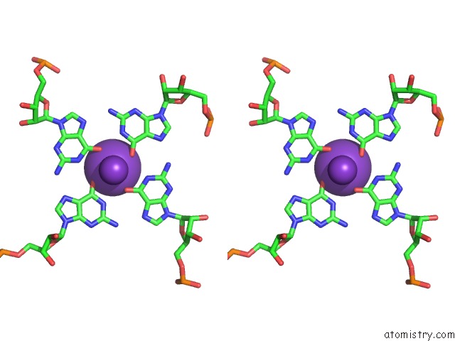
 Stereo pair view
|
A full contact list of Potassium with other atoms in the K binding
site number 3 of Crystal Structure of the Mango-II-A22U Fluorescent Aptamer Bound to TO1-Biotin within 5.0Å range:
probe
|
atom
|
residue
|
distance (Å)
|
B
|
Occ
|
A:K104
b:47.7
occ:0.50
|
O6
|
A:G10
|
2.7
|
30.8
|
1.0
|
O6
|
A:G20
|
2.8
|
33.5
|
1.0
|
O6
|
A:G26
|
2.8
|
39.5
|
1.0
|
O6
|
A:G15
|
3.0
|
35.4
|
1.0
|
K
|
A:K103
|
3.3
|
33.9
|
1.0
|
C6
|
A:G26
|
3.5
|
33.2
|
1.0
|
C6
|
A:G10
|
3.6
|
30.9
|
1.0
|
C6
|
A:G20
|
3.6
|
36.8
|
1.0
|
C6
|
A:G15
|
3.7
|
31.5
|
1.0
|
N1
|
A:G26
|
3.8
|
33.4
|
1.0
|
N1
|
A:G15
|
3.8
|
28.4
|
1.0
|
N1
|
A:G10
|
3.9
|
28.2
|
1.0
|
N1
|
A:G20
|
3.9
|
38.9
|
1.0
|
C5
|
A:G26
|
4.7
|
33.1
|
1.0
|
C5
|
A:G20
|
4.8
|
37.0
|
1.0
|
C5
|
A:G10
|
4.9
|
30.6
|
1.0
|
C5
|
A:G15
|
4.9
|
31.4
|
1.0
|
C2
|
A:G26
|
5.0
|
30.9
|
1.0
|
|
Potassium binding site 4 out
of 8 in 6c65
Go back to
Potassium Binding Sites List in 6c65
Potassium binding site 4 out
of 8 in the Crystal Structure of the Mango-II-A22U Fluorescent Aptamer Bound to TO1-Biotin
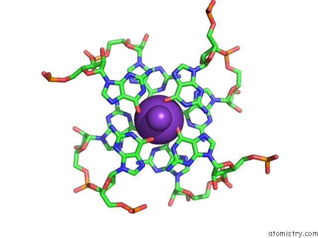
 Mono view
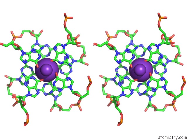
 Stereo pair view
|
A full contact list of Potassium with other atoms in the K binding
site number 4 of Crystal Structure of the Mango-II-A22U Fluorescent Aptamer Bound to TO1-Biotin within 5.0Å range:
probe
|
atom
|
residue
|
distance (Å)
|
B
|
Occ
|
B:K102
b:43.0
occ:1.00
|
O6
|
B:G15
|
2.6
|
48.6
|
1.0
|
O6
|
B:G11
|
2.7
|
46.6
|
1.0
|
O6
|
B:G16
|
2.7
|
41.0
|
1.0
|
O6
|
B:G21
|
2.7
|
49.1
|
1.0
|
O6
|
B:G27
|
2.8
|
45.8
|
1.0
|
O6
|
B:G26
|
2.9
|
50.1
|
1.0
|
O6
|
B:G10
|
2.9
|
47.0
|
1.0
|
O6
|
B:G20
|
2.9
|
43.7
|
1.0
|
K
|
B:K103
|
3.2
|
47.7
|
1.0
|
K
|
B:K104
|
3.4
|
53.7
|
1.0
|
C6
|
B:G15
|
3.5
|
48.2
|
1.0
|
C6
|
B:G10
|
3.5
|
45.7
|
1.0
|
C6
|
B:G16
|
3.5
|
44.1
|
1.0
|
C6
|
B:G20
|
3.6
|
46.4
|
1.0
|
C6
|
B:G21
|
3.6
|
48.4
|
1.0
|
C6
|
B:G11
|
3.6
|
45.4
|
1.0
|
C6
|
B:G27
|
3.7
|
43.1
|
1.0
|
C6
|
B:G26
|
3.7
|
45.6
|
1.0
|
N1
|
B:G10
|
3.7
|
44.4
|
1.0
|
N1
|
B:G20
|
3.8
|
48.0
|
1.0
|
N1
|
B:G21
|
3.8
|
47.6
|
1.0
|
N1
|
B:G15
|
3.8
|
44.5
|
1.0
|
N1
|
B:G16
|
3.9
|
44.2
|
1.0
|
N1
|
B:G26
|
3.9
|
41.7
|
1.0
|
N1
|
B:G11
|
3.9
|
47.6
|
1.0
|
N1
|
B:G27
|
4.0
|
41.5
|
1.0
|
C5
|
B:G10
|
4.7
|
45.8
|
1.0
|
C5
|
B:G20
|
4.7
|
45.7
|
1.0
|
C5
|
B:G15
|
4.8
|
49.0
|
1.0
|
C5
|
B:G16
|
4.8
|
46.2
|
1.0
|
C5
|
B:G11
|
4.8
|
41.8
|
1.0
|
C5
|
B:G21
|
4.8
|
48.9
|
1.0
|
C2
|
B:G10
|
4.9
|
44.1
|
1.0
|
C5
|
B:G27
|
4.9
|
40.1
|
1.0
|
C2
|
B:G20
|
4.9
|
47.7
|
1.0
|
C5
|
B:G26
|
5.0
|
44.3
|
1.0
|
|
Potassium binding site 5 out
of 8 in 6c65
Go back to
Potassium Binding Sites List in 6c65
Potassium binding site 5 out
of 8 in the Crystal Structure of the Mango-II-A22U Fluorescent Aptamer Bound to TO1-Biotin
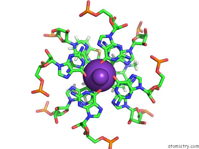
 Mono view
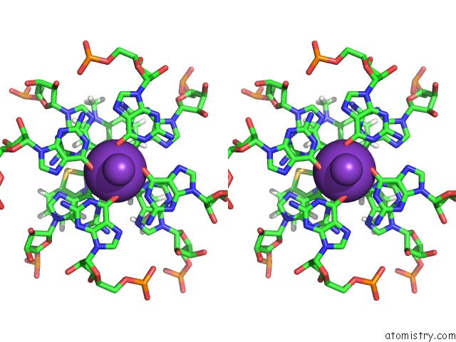
 Stereo pair view
|
A full contact list of Potassium with other atoms in the K binding
site number 5 of Crystal Structure of the Mango-II-A22U Fluorescent Aptamer Bound to TO1-Biotin within 5.0Å range:
probe
|
atom
|
residue
|
distance (Å)
|
B
|
Occ
|
B:K103
b:47.7
occ:1.00
|
O6
|
B:G11
|
2.6
|
46.6
|
1.0
|
O6
|
B:G27
|
2.6
|
45.8
|
1.0
|
O6
|
B:G24
|
2.7
|
51.0
|
1.0
|
O6
|
B:G13
|
2.7
|
50.2
|
1.0
|
O6
|
B:G21
|
2.8
|
49.1
|
1.0
|
O6
|
B:G16
|
2.8
|
41.0
|
1.0
|
O6
|
B:G18
|
2.9
|
48.5
|
1.0
|
O6
|
B:G29
|
3.0
|
46.5
|
1.0
|
K
|
B:K102
|
3.2
|
43.0
|
1.0
|
C6
|
B:G27
|
3.4
|
43.1
|
1.0
|
C6
|
B:G24
|
3.5
|
53.0
|
1.0
|
C6
|
B:G11
|
3.5
|
45.4
|
1.0
|
C6
|
B:G21
|
3.5
|
48.4
|
1.0
|
C6
|
B:G13
|
3.6
|
48.6
|
1.0
|
C6
|
B:G16
|
3.6
|
44.1
|
1.0
|
N1
|
B:G27
|
3.7
|
41.5
|
1.0
|
N1
|
B:G21
|
3.7
|
47.6
|
1.0
|
C6
|
B:G29
|
3.7
|
45.5
|
1.0
|
N1
|
B:G24
|
3.8
|
53.5
|
1.0
|
C6
|
B:G18
|
3.9
|
47.4
|
1.0
|
N1
|
B:G11
|
3.9
|
47.6
|
1.0
|
N1
|
B:G16
|
3.9
|
44.2
|
1.0
|
N1
|
B:G13
|
4.0
|
45.7
|
1.0
|
N1
|
B:G29
|
4.0
|
45.6
|
1.0
|
N1
|
B:G18
|
4.4
|
47.6
|
1.0
|
H111
|
B:EKJ101
|
4.7
|
70.2
|
1.0
|
C5
|
B:G24
|
4.7
|
53.5
|
1.0
|
C5
|
B:G11
|
4.7
|
41.8
|
1.0
|
C5
|
B:G27
|
4.7
|
40.1
|
1.0
|
C5
|
B:G13
|
4.8
|
48.0
|
1.0
|
C5
|
B:G21
|
4.8
|
48.9
|
1.0
|
C5
|
B:G29
|
4.9
|
44.7
|
1.0
|
C5
|
B:G16
|
4.9
|
46.2
|
1.0
|
C2
|
B:G27
|
4.9
|
43.0
|
1.0
|
C2
|
B:G21
|
4.9
|
49.7
|
1.0
|
C5
|
B:G18
|
5.0
|
48.8
|
1.0
|
C2
|
B:G24
|
5.0
|
55.9
|
1.0
|
|
Potassium binding site 6 out
of 8 in 6c65
Go back to
Potassium Binding Sites List in 6c65
Potassium binding site 6 out
of 8 in the Crystal Structure of the Mango-II-A22U Fluorescent Aptamer Bound to TO1-Biotin
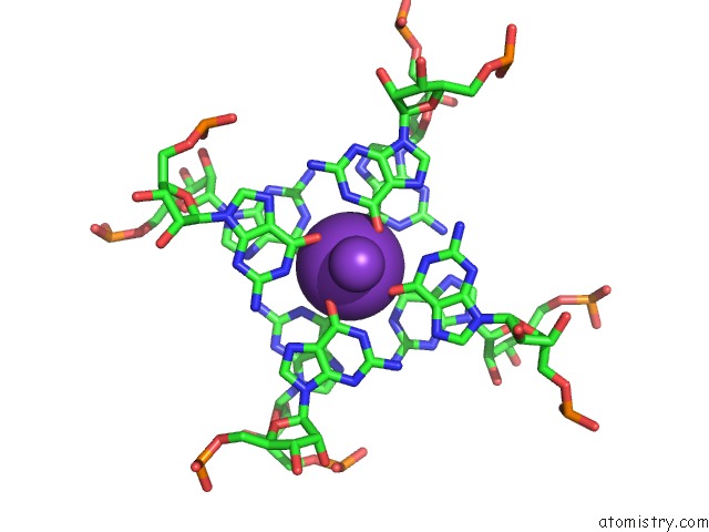
 Mono view
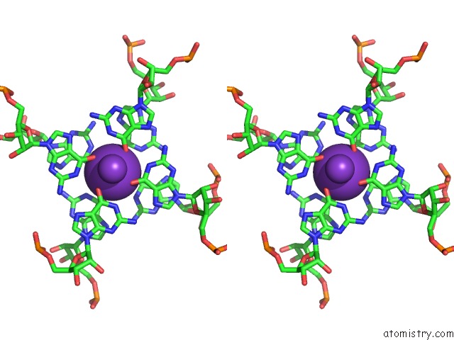
 Stereo pair view
|
A full contact list of Potassium with other atoms in the K binding
site number 6 of Crystal Structure of the Mango-II-A22U Fluorescent Aptamer Bound to TO1-Biotin within 5.0Å range:
probe
|
atom
|
residue
|
distance (Å)
|
B
|
Occ
|
B:K104
b:53.7
occ:1.00
|
O6
|
B:G10
|
2.6
|
47.0
|
1.0
|
O6
|
C:G26
|
2.7
|
50.1
|
1.0
|
O6
|
C:G15
|
2.8
|
47.9
|
1.0
|
O6
|
C:G10
|
2.8
|
49.1
|
1.0
|
O6
|
B:G20
|
2.8
|
43.7
|
1.0
|
O6
|
C:G20
|
2.8
|
47.0
|
1.0
|
O6
|
B:G15
|
2.9
|
48.6
|
1.0
|
O6
|
B:G26
|
3.0
|
50.1
|
1.0
|
K
|
C:K102
|
3.3
|
40.1
|
1.0
|
K
|
B:K102
|
3.4
|
43.0
|
1.0
|
C6
|
B:G10
|
3.4
|
45.7
|
1.0
|
C6
|
C:G26
|
3.5
|
49.1
|
1.0
|
C6
|
C:G20
|
3.5
|
46.3
|
1.0
|
C6
|
C:G15
|
3.6
|
47.6
|
1.0
|
N1
|
B:G10
|
3.7
|
44.4
|
1.0
|
C6
|
B:G20
|
3.7
|
46.4
|
1.0
|
N1
|
C:G20
|
3.7
|
48.5
|
1.0
|
C6
|
B:G26
|
3.7
|
45.6
|
1.0
|
C6
|
B:G15
|
3.7
|
48.2
|
1.0
|
C6
|
C:G10
|
3.8
|
47.9
|
1.0
|
N1
|
B:G20
|
3.8
|
48.0
|
1.0
|
N1
|
C:G15
|
3.8
|
46.3
|
1.0
|
N1
|
C:G26
|
3.9
|
48.7
|
1.0
|
N1
|
B:G26
|
3.9
|
41.7
|
1.0
|
N1
|
B:G15
|
4.0
|
44.5
|
1.0
|
N1
|
C:G10
|
4.1
|
47.5
|
1.0
|
C5
|
C:G26
|
4.6
|
48.5
|
1.0
|
C5
|
B:G10
|
4.7
|
45.8
|
1.0
|
C5
|
C:G15
|
4.8
|
47.4
|
1.0
|
C5
|
C:G20
|
4.8
|
46.2
|
1.0
|
C5
|
B:G15
|
4.8
|
49.0
|
1.0
|
C5
|
B:G26
|
4.9
|
44.3
|
1.0
|
C2
|
C:G20
|
4.9
|
47.5
|
1.0
|
C2
|
B:G10
|
4.9
|
44.1
|
1.0
|
C5
|
B:G20
|
5.0
|
45.7
|
1.0
|
|
Potassium binding site 7 out
of 8 in 6c65
Go back to
Potassium Binding Sites List in 6c65
Potassium binding site 7 out
of 8 in the Crystal Structure of the Mango-II-A22U Fluorescent Aptamer Bound to TO1-Biotin
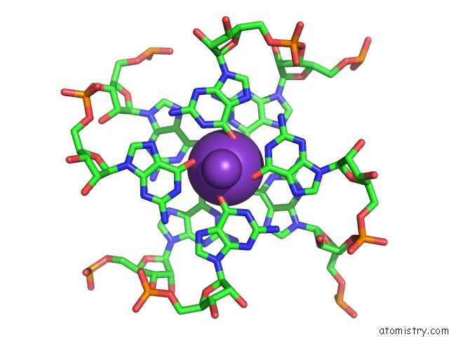
 Mono view
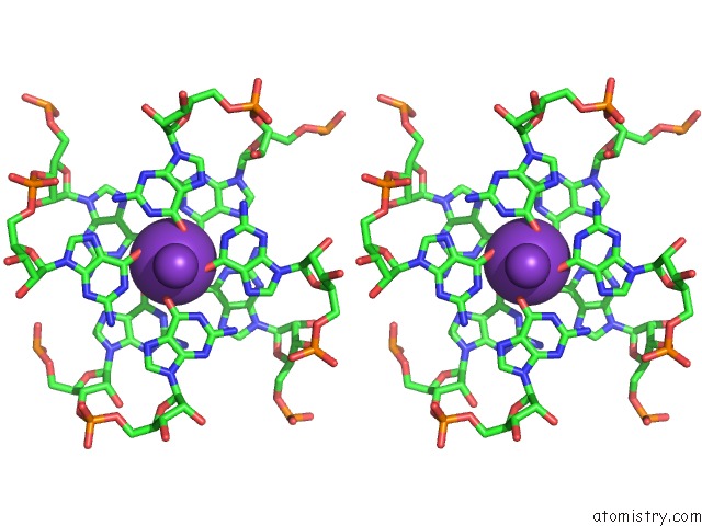
 Stereo pair view
|
A full contact list of Potassium with other atoms in the K binding
site number 7 of Crystal Structure of the Mango-II-A22U Fluorescent Aptamer Bound to TO1-Biotin within 5.0Å range:
probe
|
atom
|
residue
|
distance (Å)
|
B
|
Occ
|
C:K102
b:40.1
occ:1.00
|
O6
|
C:G27
|
2.6
|
50.9
|
1.0
|
O6
|
C:G16
|
2.7
|
44.7
|
1.0
|
O6
|
C:G15
|
2.7
|
47.9
|
1.0
|
O6
|
C:G11
|
2.7
|
50.4
|
1.0
|
O6
|
C:G26
|
2.7
|
50.1
|
1.0
|
O6
|
C:G20
|
2.8
|
47.0
|
1.0
|
O6
|
C:G21
|
2.9
|
49.7
|
1.0
|
O6
|
C:G10
|
2.9
|
49.1
|
1.0
|
K
|
B:K104
|
3.3
|
53.7
|
1.0
|
K
|
C:K103
|
3.4
|
57.8
|
1.0
|
C6
|
C:G16
|
3.5
|
43.6
|
1.0
|
C6
|
C:G27
|
3.5
|
51.9
|
1.0
|
C6
|
C:G11
|
3.5
|
51.7
|
1.0
|
C6
|
C:G15
|
3.5
|
47.6
|
1.0
|
C6
|
C:G10
|
3.6
|
47.9
|
1.0
|
C6
|
C:G26
|
3.6
|
49.1
|
1.0
|
C6
|
C:G20
|
3.6
|
46.3
|
1.0
|
N1
|
C:G11
|
3.7
|
49.0
|
1.0
|
C6
|
C:G21
|
3.8
|
49.5
|
1.0
|
N1
|
C:G16
|
3.8
|
45.1
|
1.0
|
N1
|
C:G26
|
3.8
|
48.7
|
1.0
|
N1
|
C:G10
|
3.8
|
47.5
|
1.0
|
N1
|
C:G20
|
3.8
|
48.5
|
1.0
|
N1
|
C:G15
|
3.9
|
46.3
|
1.0
|
N1
|
C:G27
|
3.9
|
50.5
|
1.0
|
N1
|
C:G21
|
4.0
|
51.6
|
1.0
|
C5
|
C:G27
|
4.6
|
48.4
|
1.0
|
C5
|
C:G16
|
4.7
|
43.8
|
1.0
|
C5
|
C:G10
|
4.7
|
47.6
|
1.0
|
C5
|
C:G11
|
4.7
|
52.6
|
1.0
|
C5
|
C:G15
|
4.7
|
47.4
|
1.0
|
C5
|
C:G20
|
4.8
|
46.2
|
1.0
|
C5
|
C:G26
|
4.9
|
48.5
|
1.0
|
C5
|
C:G21
|
5.0
|
51.3
|
1.0
|
C2
|
C:G11
|
5.0
|
50.3
|
1.0
|
|
Potassium binding site 8 out
of 8 in 6c65
Go back to
Potassium Binding Sites List in 6c65
Potassium binding site 8 out
of 8 in the Crystal Structure of the Mango-II-A22U Fluorescent Aptamer Bound to TO1-Biotin
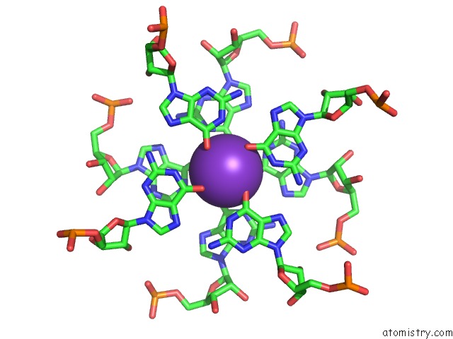
 Mono view
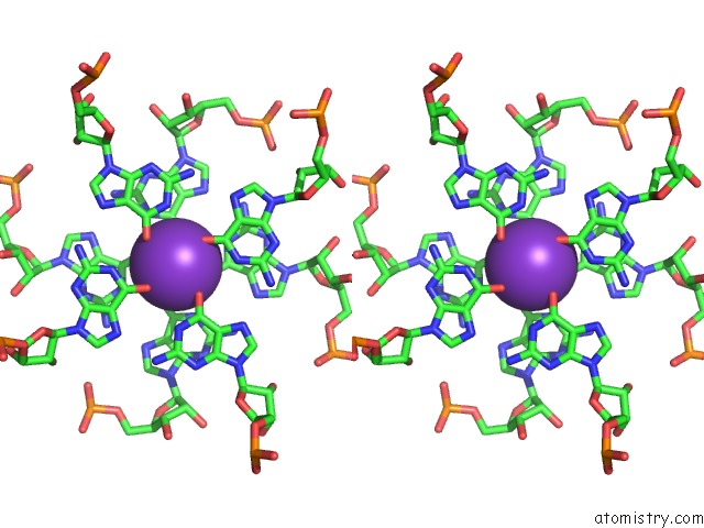
 Stereo pair view
|
A full contact list of Potassium with other atoms in the K binding
site number 8 of Crystal Structure of the Mango-II-A22U Fluorescent Aptamer Bound to TO1-Biotin within 5.0Å range:
probe
|
atom
|
residue
|
distance (Å)
|
B
|
Occ
|
C:K103
b:57.8
occ:1.00
|
O6
|
C:G27
|
2.5
|
50.9
|
1.0
|
O6
|
C:G24
|
2.7
|
55.7
|
1.0
|
O6
|
C:G21
|
2.7
|
49.7
|
1.0
|
O6
|
C:G18
|
2.8
|
56.3
|
1.0
|
O6
|
C:G11
|
2.8
|
50.4
|
1.0
|
O6
|
C:G13
|
2.8
|
54.2
|
1.0
|
O6
|
C:G29
|
2.9
|
56.3
|
1.0
|
O6
|
C:G16
|
3.0
|
44.7
|
1.0
|
C6
|
C:G27
|
3.4
|
51.9
|
1.0
|
K
|
C:K102
|
3.4
|
40.1
|
1.0
|
C6
|
C:G24
|
3.6
|
56.5
|
1.0
|
C6
|
C:G21
|
3.6
|
49.5
|
1.0
|
C6
|
C:G16
|
3.7
|
43.6
|
1.0
|
C6
|
C:G29
|
3.7
|
54.6
|
1.0
|
C6
|
C:G11
|
3.7
|
51.7
|
1.0
|
C6
|
C:G18
|
3.7
|
51.4
|
1.0
|
C6
|
C:G13
|
3.7
|
56.1
|
1.0
|
N1
|
C:G16
|
3.8
|
45.1
|
1.0
|
N1
|
C:G29
|
3.8
|
53.0
|
1.0
|
N1
|
C:G27
|
3.9
|
50.5
|
1.0
|
N1
|
C:G21
|
3.9
|
51.6
|
1.0
|
N1
|
C:G11
|
3.9
|
49.0
|
1.0
|
N1
|
C:G24
|
3.9
|
55.2
|
1.0
|
N1
|
C:G18
|
4.0
|
46.3
|
1.0
|
N1
|
C:G13
|
4.1
|
53.1
|
1.0
|
C5
|
C:G27
|
4.6
|
48.4
|
1.0
|
C5
|
C:G24
|
4.7
|
58.8
|
1.0
|
C5
|
C:G21
|
4.8
|
51.3
|
1.0
|
C5
|
C:G18
|
4.9
|
52.3
|
1.0
|
C5
|
C:G16
|
4.9
|
43.8
|
1.0
|
C5
|
C:G13
|
4.9
|
56.5
|
1.0
|
C5
|
C:G29
|
4.9
|
56.4
|
1.0
|
C5
|
C:G11
|
4.9
|
52.6
|
1.0
|
C2
|
C:G16
|
5.0
|
46.7
|
1.0
|
|
Reference:
R.J.Trachman 3Rd.,
A.Abdolahzadeh,
A.Andreoni,
R.Cojocaru,
J.R.Knutson,
M.Ryckelynck,
P.J.Unrau,
A.R.Ferre-D'amare.
Crystal Structures of the Mango-II Rna Aptamer Reveal Heterogeneous Fluorophore Binding and Guide Engineering of Variants with Improved Selectivity and Brightness. Biochemistry V. 57 3544 2018.
ISSN: ISSN 1520-4995
PubMed: 29768001
DOI: 10.1021/ACS.BIOCHEM.8B00399
Page generated: Mon Aug 12 15:30:45 2024
|