Potassium »
PDB 6bd3-6csr »
6c63 »
Potassium in PDB 6c63: Crystal Structure of the Mango-II Fluorescent Aptamer Bound to TO1- Biotin
Protein crystallography data
The structure of Crystal Structure of the Mango-II Fluorescent Aptamer Bound to TO1- Biotin, PDB code: 6c63
was solved by
R.J.Trachman,
A.R.Ferre-D'amare,
with X-Ray Crystallography technique. A brief refinement statistics is given in the table below:
| Resolution Low / High (Å) | 91.21 / 2.90 |
| Space group | C 2 2 21 |
| Cell size a, b, c (Å), α, β, γ (°) | 36.831, 182.412, 107.494, 90.00, 90.00, 90.00 |
| R / Rfree (%) | 18.5 / 23 |
Potassium Binding Sites:
The binding sites of Potassium atom in the Crystal Structure of the Mango-II Fluorescent Aptamer Bound to TO1- Biotin
(pdb code 6c63). This binding sites where shown within
5.0 Angstroms radius around Potassium atom.
In total 8 binding sites of Potassium where determined in the Crystal Structure of the Mango-II Fluorescent Aptamer Bound to TO1- Biotin, PDB code: 6c63:
Jump to Potassium binding site number: 1; 2; 3; 4; 5; 6; 7; 8;
In total 8 binding sites of Potassium where determined in the Crystal Structure of the Mango-II Fluorescent Aptamer Bound to TO1- Biotin, PDB code: 6c63:
Jump to Potassium binding site number: 1; 2; 3; 4; 5; 6; 7; 8;
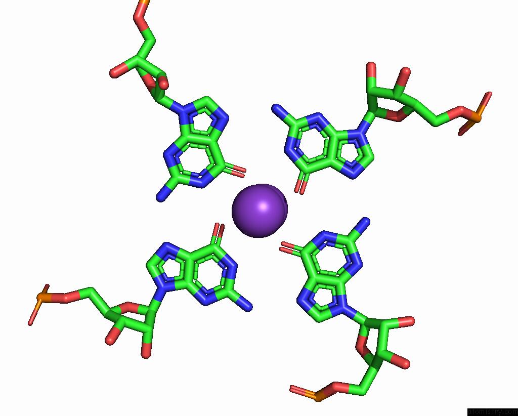
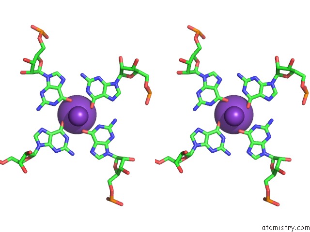
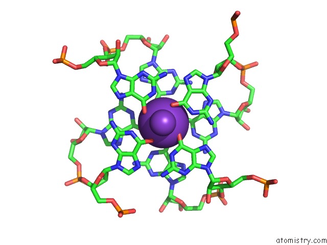
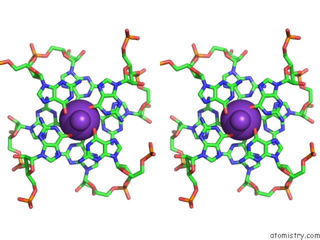
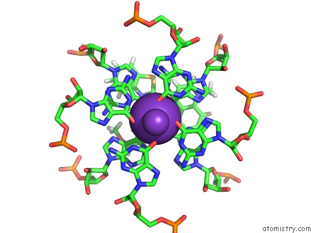
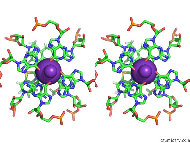
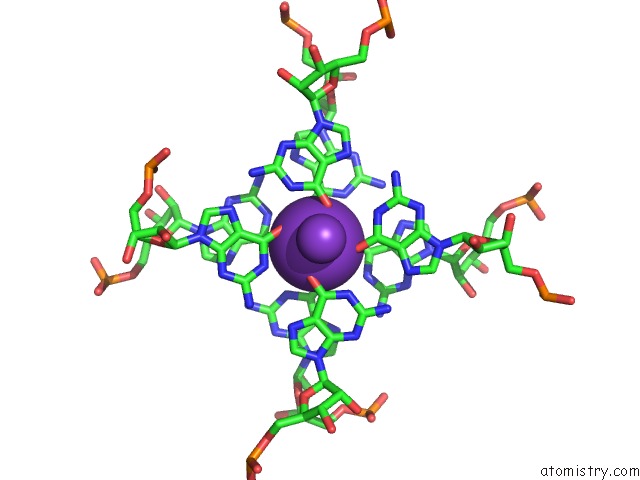
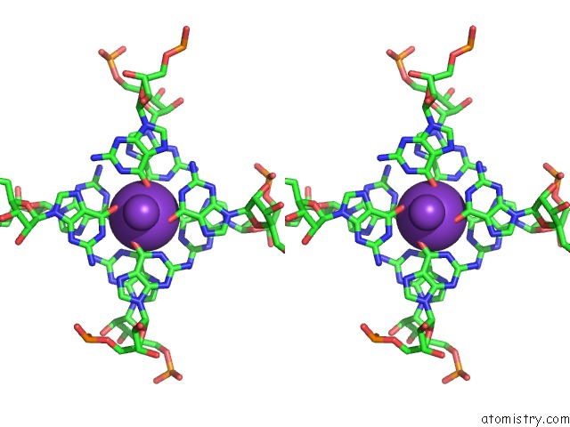
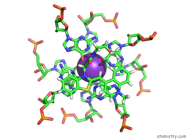
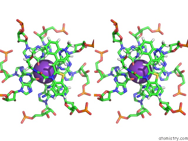
Potassium binding site 1 out of 8 in 6c63
Go back to
Potassium binding site 1 out
of 8 in the Crystal Structure of the Mango-II Fluorescent Aptamer Bound to TO1- Biotin
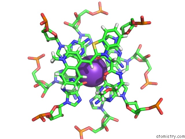
Mono view
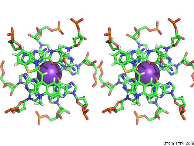
Stereo pair view
Mono view
Stereo pair view
A full contact list of Potassium with other atoms in the K binding
site number 1 of Crystal Structure of the Mango-II Fluorescent Aptamer Bound to TO1- Biotin within 5.0Å range:
|
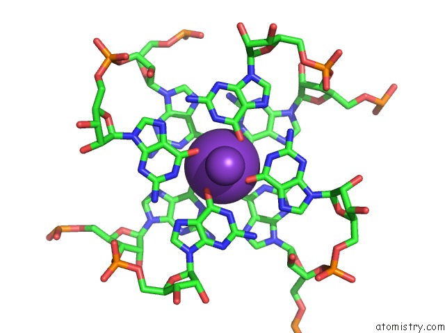
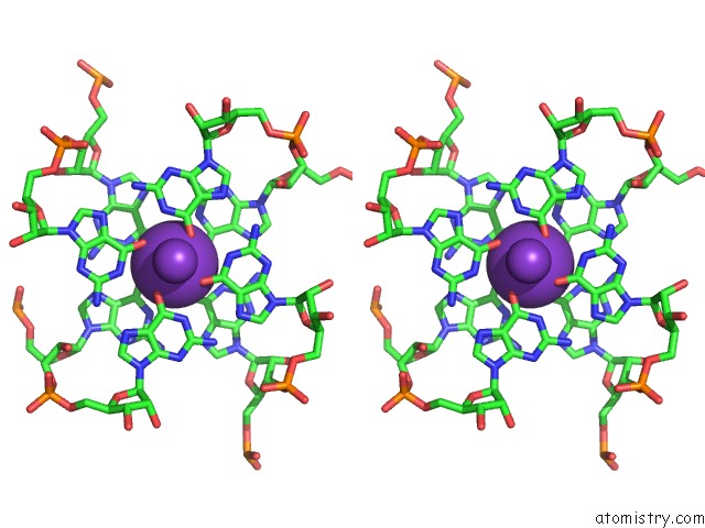
Potassium binding site 2 out of 8 in 6c63
Go back to
Potassium binding site 2 out
of 8 in the Crystal Structure of the Mango-II Fluorescent Aptamer Bound to TO1- Biotin
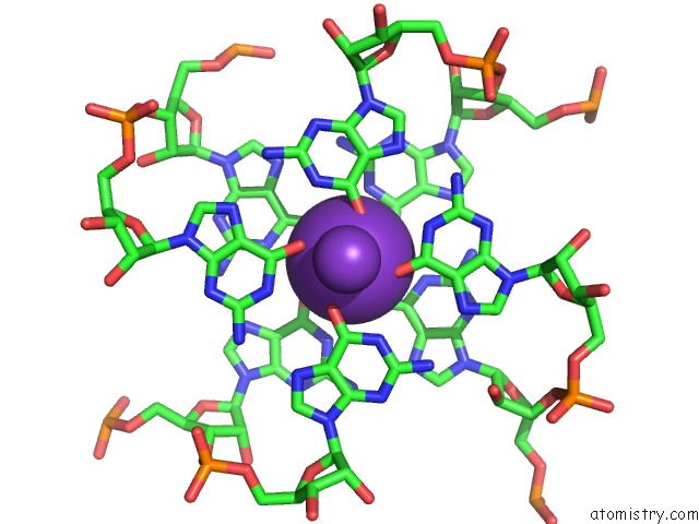
Mono view
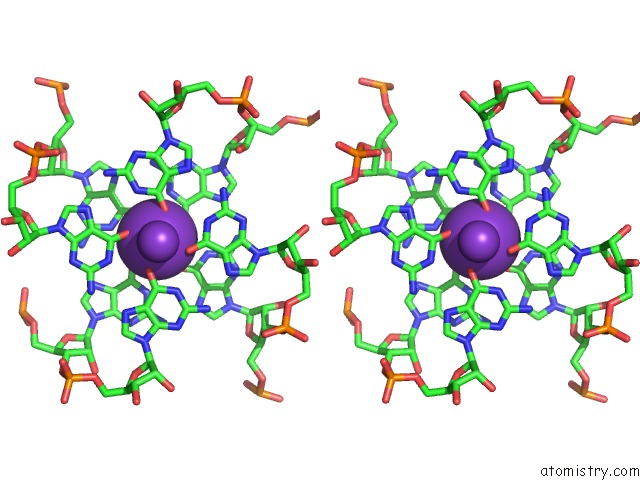
Stereo pair view
Mono view
Stereo pair view
A full contact list of Potassium with other atoms in the K binding
site number 2 of Crystal Structure of the Mango-II Fluorescent Aptamer Bound to TO1- Biotin within 5.0Å range:
|
Potassium binding site 3 out of 8 in 6c63
Go back to
Potassium binding site 3 out
of 8 in the Crystal Structure of the Mango-II Fluorescent Aptamer Bound to TO1- Biotin
Mono view
Stereo pair view
Mono view
Stereo pair view
A full contact list of Potassium with other atoms in the K binding
site number 3 of Crystal Structure of the Mango-II Fluorescent Aptamer Bound to TO1- Biotin within 5.0Å range:
|
Potassium binding site 4 out of 8 in 6c63
Go back to
Potassium binding site 4 out
of 8 in the Crystal Structure of the Mango-II Fluorescent Aptamer Bound to TO1- Biotin
Mono view
Stereo pair view
Mono view
Stereo pair view
A full contact list of Potassium with other atoms in the K binding
site number 4 of Crystal Structure of the Mango-II Fluorescent Aptamer Bound to TO1- Biotin within 5.0Å range:
|
Potassium binding site 5 out of 8 in 6c63
Go back to
Potassium binding site 5 out
of 8 in the Crystal Structure of the Mango-II Fluorescent Aptamer Bound to TO1- Biotin
Mono view
Stereo pair view
Mono view
Stereo pair view
A full contact list of Potassium with other atoms in the K binding
site number 5 of Crystal Structure of the Mango-II Fluorescent Aptamer Bound to TO1- Biotin within 5.0Å range:
|
Potassium binding site 6 out of 8 in 6c63
Go back to
Potassium binding site 6 out
of 8 in the Crystal Structure of the Mango-II Fluorescent Aptamer Bound to TO1- Biotin
Mono view
Stereo pair view
Mono view
Stereo pair view
A full contact list of Potassium with other atoms in the K binding
site number 6 of Crystal Structure of the Mango-II Fluorescent Aptamer Bound to TO1- Biotin within 5.0Å range:
|
Potassium binding site 7 out of 8 in 6c63
Go back to
Potassium binding site 7 out
of 8 in the Crystal Structure of the Mango-II Fluorescent Aptamer Bound to TO1- Biotin
Mono view
Stereo pair view
Mono view
Stereo pair view
A full contact list of Potassium with other atoms in the K binding
site number 7 of Crystal Structure of the Mango-II Fluorescent Aptamer Bound to TO1- Biotin within 5.0Å range:
|
Potassium binding site 8 out of 8 in 6c63
Go back to
Potassium binding site 8 out
of 8 in the Crystal Structure of the Mango-II Fluorescent Aptamer Bound to TO1- Biotin
Mono view
Stereo pair view
Mono view
Stereo pair view
A full contact list of Potassium with other atoms in the K binding
site number 8 of Crystal Structure of the Mango-II Fluorescent Aptamer Bound to TO1- Biotin within 5.0Å range:
|
Reference:
R.J.Trachman 3Rd.,
A.Abdolahzadeh,
A.Andreoni,
R.Cojocaru,
J.R.Knutson,
M.Ryckelynck,
P.J.Unrau,
A.R.Ferre-D'amare.
Crystal Structures of the Mango-II Rna Aptamer Reveal Heterogeneous Fluorophore Binding and Guide Engineering of Variants with Improved Selectivity and Brightness. Biochemistry V. 57 3544 2018.
ISSN: ISSN 1520-4995
PubMed: 29768001
DOI: 10.1021/ACS.BIOCHEM.8B00399
Page generated: Mon Aug 12 15:30:31 2024
ISSN: ISSN 1520-4995
PubMed: 29768001
DOI: 10.1021/ACS.BIOCHEM.8B00399
Last articles
Zn in 9J0NZn in 9J0O
Zn in 9J0P
Zn in 9FJX
Zn in 9EKB
Zn in 9C0F
Zn in 9CAH
Zn in 9CH0
Zn in 9CH3
Zn in 9CH1