Potassium »
PDB 5ksd-5mq0 »
5lwo »
Potassium in PDB 5lwo: Structure of Spin-Labelled T4 Lysozyme Mutant L115C-R119C-R1 at 100K
Enzymatic activity of Structure of Spin-Labelled T4 Lysozyme Mutant L115C-R119C-R1 at 100K
All present enzymatic activity of Structure of Spin-Labelled T4 Lysozyme Mutant L115C-R119C-R1 at 100K:
3.2.1.17;
3.2.1.17;
Protein crystallography data
The structure of Structure of Spin-Labelled T4 Lysozyme Mutant L115C-R119C-R1 at 100K, PDB code: 5lwo
was solved by
B.Loll,
P.Consentius,
U.Gohlke,
R.Mueller,
M.Kaupp,
U.Heinemann,
M.C.Wahl,
T.Risse,
with X-Ray Crystallography technique. A brief refinement statistics is given in the table below:
| Resolution Low / High (Å) | 18.30 / 1.18 |
| Space group | P 32 2 1 |
| Cell size a, b, c (Å), α, β, γ (°) | 60.170, 60.170, 97.720, 90.00, 90.00, 120.00 |
| R / Rfree (%) | 14.1 / 16.4 |
Other elements in 5lwo:
The structure of Structure of Spin-Labelled T4 Lysozyme Mutant L115C-R119C-R1 at 100K also contains other interesting chemical elements:
| Chlorine | (Cl) | 4 atoms |
Potassium Binding Sites:
The binding sites of Potassium atom in the Structure of Spin-Labelled T4 Lysozyme Mutant L115C-R119C-R1 at 100K
(pdb code 5lwo). This binding sites where shown within
5.0 Angstroms radius around Potassium atom.
In total 2 binding sites of Potassium where determined in the Structure of Spin-Labelled T4 Lysozyme Mutant L115C-R119C-R1 at 100K, PDB code: 5lwo:
Jump to Potassium binding site number: 1; 2;
In total 2 binding sites of Potassium where determined in the Structure of Spin-Labelled T4 Lysozyme Mutant L115C-R119C-R1 at 100K, PDB code: 5lwo:
Jump to Potassium binding site number: 1; 2;
Potassium binding site 1 out of 2 in 5lwo
Go back to
Potassium binding site 1 out
of 2 in the Structure of Spin-Labelled T4 Lysozyme Mutant L115C-R119C-R1 at 100K

Mono view
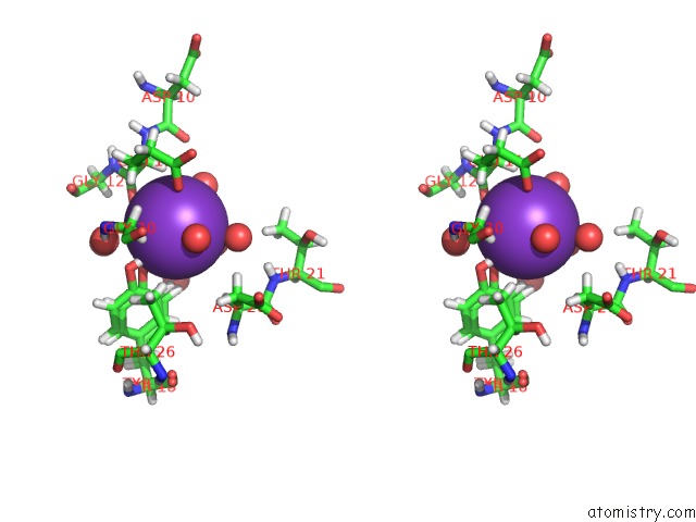
Stereo pair view
Mono view
Stereo pair view
A full contact list of Potassium with other atoms in the K binding
site number 1 of Structure of Spin-Labelled T4 Lysozyme Mutant L115C-R119C-R1 at 100K within 5.0Å range:
|
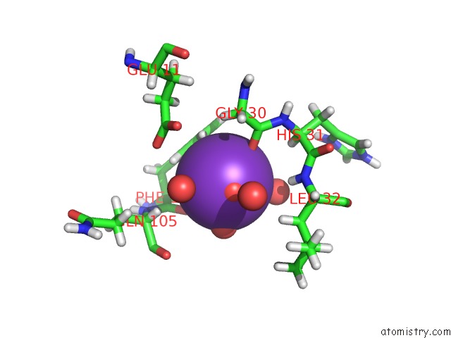
Potassium binding site 2 out of 2 in 5lwo
Go back to
Potassium binding site 2 out
of 2 in the Structure of Spin-Labelled T4 Lysozyme Mutant L115C-R119C-R1 at 100K
Mono view

Stereo pair view
Mono view
Stereo pair view
A full contact list of Potassium with other atoms in the K binding
site number 2 of Structure of Spin-Labelled T4 Lysozyme Mutant L115C-R119C-R1 at 100K within 5.0Å range:
|
Reference:
P.Consentius,
B.Loll,
U.Gohlke,
C.Alings,
C.Muller,
R.Muller,
C.Teutloff,
U.Heinemann,
M.Kaupp,
M.C.Wahl,
T.Risse.
Internal Dynamics of the 3-Pyrroline-N-Oxide Ring in Spin-Labeled Proteins. J Phys Chem Lett V. 8 1113 2017.
ISSN: ESSN 1948-7185
PubMed: 28221042
DOI: 10.1021/ACS.JPCLETT.6B02971
Page generated: Sat Aug 9 09:32:36 2025
ISSN: ESSN 1948-7185
PubMed: 28221042
DOI: 10.1021/ACS.JPCLETT.6B02971
Last articles
K in 7Q3XK in 7QDN
K in 7QF6
K in 7Q0G
K in 7Q1C
K in 7Q1B
K in 7PXH
K in 7PZJ
K in 7PXG
K in 7PWY