Potassium »
PDB 5g17-5imu »
5hnk »
Potassium in PDB 5hnk: Crystal Structure of T5FEN in Complex Intact Substrate and Metal Ions.
Enzymatic activity of Crystal Structure of T5FEN in Complex Intact Substrate and Metal Ions.
All present enzymatic activity of Crystal Structure of T5FEN in Complex Intact Substrate and Metal Ions.:
3.1.11.3;
3.1.11.3;
Protein crystallography data
The structure of Crystal Structure of T5FEN in Complex Intact Substrate and Metal Ions., PDB code: 5hnk
was solved by
F.A.Almalki,
M.Feng,
J.Zhang,
S.E.Sedelnikova,
J.B.Rafferty,
J.R.Sayers,
P.J.Artymiuk,
with X-Ray Crystallography technique. A brief refinement statistics is given in the table below:
| Resolution Low / High (Å) | 42.19 / 2.22 |
| Space group | P 21 21 21 |
| Cell size a, b, c (Å), α, β, γ (°) | 44.720, 109.940, 127.340, 90.00, 90.00, 90.00 |
| R / Rfree (%) | 18.3 / 23.4 |
Other elements in 5hnk:
The structure of Crystal Structure of T5FEN in Complex Intact Substrate and Metal Ions. also contains other interesting chemical elements:
| Magnesium | (Mg) | 2 atoms |
Potassium Binding Sites:
The binding sites of Potassium atom in the Crystal Structure of T5FEN in Complex Intact Substrate and Metal Ions.
(pdb code 5hnk). This binding sites where shown within
5.0 Angstroms radius around Potassium atom.
In total only one binding site of Potassium was determined in the Crystal Structure of T5FEN in Complex Intact Substrate and Metal Ions., PDB code: 5hnk:
In total only one binding site of Potassium was determined in the Crystal Structure of T5FEN in Complex Intact Substrate and Metal Ions., PDB code: 5hnk:
Potassium binding site 1 out of 1 in 5hnk
Go back to
Potassium binding site 1 out
of 1 in the Crystal Structure of T5FEN in Complex Intact Substrate and Metal Ions.
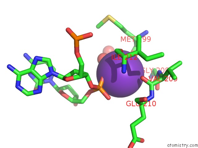
Mono view
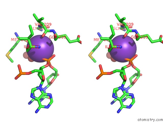
Stereo pair view
Mono view
Stereo pair view
A full contact list of Potassium with other atoms in the K binding
site number 1 of Crystal Structure of T5FEN in Complex Intact Substrate and Metal Ions. within 5.0Å range:
|
Reference:
F.A.Almalki,
C.S.Flemming,
J.Zhang,
M.Feng,
S.E.Sedelnikova,
T.Ceska,
J.B.Rafferty,
J.R.Sayers,
P.J.Artymiuk.
Direct Observation of Dna Threading in Flap Endonuclease Complexes. Nat.Struct.Mol.Biol. V. 23 640 2016.
ISSN: ESSN 1545-9985
PubMed: 27273516
DOI: 10.1038/NSMB.3241
Page generated: Sat Aug 9 09:14:05 2025
ISSN: ESSN 1545-9985
PubMed: 27273516
DOI: 10.1038/NSMB.3241
Last articles
K in 8K1EK in 8JZG
K in 8K0U
K in 8K0T
K in 8JGW
K in 8JZA
K in 8JC7
K in 8JGR
K in 8IYM
K in 8JAH