Potassium »
PDB 4cn5-4edj »
4e1v »
Potassium in PDB 4e1v: X-Ray Structure of the Uridine Phosphorylase From Salmonella Typhimurium in Complex with 5-Fluorouracil at 2.15 A Resolution
Enzymatic activity of X-Ray Structure of the Uridine Phosphorylase From Salmonella Typhimurium in Complex with 5-Fluorouracil at 2.15 A Resolution
All present enzymatic activity of X-Ray Structure of the Uridine Phosphorylase From Salmonella Typhimurium in Complex with 5-Fluorouracil at 2.15 A Resolution:
2.4.2.3;
2.4.2.3;
Protein crystallography data
The structure of X-Ray Structure of the Uridine Phosphorylase From Salmonella Typhimurium in Complex with 5-Fluorouracil at 2.15 A Resolution, PDB code: 4e1v
was solved by
A.A.Lashkov,
S.E.Sotnichenko,
I.I.Prokofev,
A.G.Gabdoulkhakov,
A.M.Mikhailov,
with X-Ray Crystallography technique. A brief refinement statistics is given in the table below:
| Resolution Low / High (Å) | 28.88 / 2.15 |
| Space group | C 1 2 1 |
| Cell size a, b, c (Å), α, β, γ (°) | 158.490, 93.210, 149.970, 90.00, 90.82, 90.00 |
| R / Rfree (%) | 19.4 / 23.8 |
Other elements in 4e1v:
The structure of X-Ray Structure of the Uridine Phosphorylase From Salmonella Typhimurium in Complex with 5-Fluorouracil at 2.15 A Resolution also contains other interesting chemical elements:
| Fluorine | (F) | 8 atoms |
Potassium Binding Sites:
The binding sites of Potassium atom in the X-Ray Structure of the Uridine Phosphorylase From Salmonella Typhimurium in Complex with 5-Fluorouracil at 2.15 A Resolution
(pdb code 4e1v). This binding sites where shown within
5.0 Angstroms radius around Potassium atom.
In total 3 binding sites of Potassium where determined in the X-Ray Structure of the Uridine Phosphorylase From Salmonella Typhimurium in Complex with 5-Fluorouracil at 2.15 A Resolution, PDB code: 4e1v:
Jump to Potassium binding site number: 1; 2; 3;
In total 3 binding sites of Potassium where determined in the X-Ray Structure of the Uridine Phosphorylase From Salmonella Typhimurium in Complex with 5-Fluorouracil at 2.15 A Resolution, PDB code: 4e1v:
Jump to Potassium binding site number: 1; 2; 3;
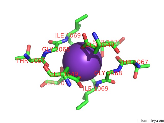
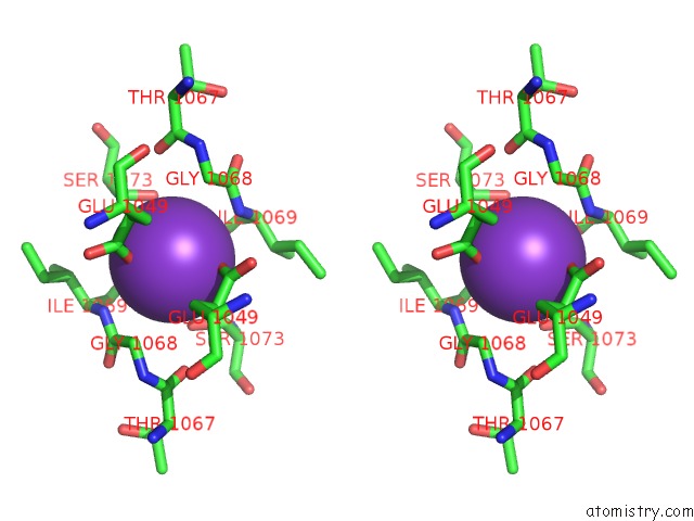
Potassium binding site 1 out of 3 in 4e1v
Go back to
Potassium binding site 1 out
of 3 in the X-Ray Structure of the Uridine Phosphorylase From Salmonella Typhimurium in Complex with 5-Fluorouracil at 2.15 A Resolution
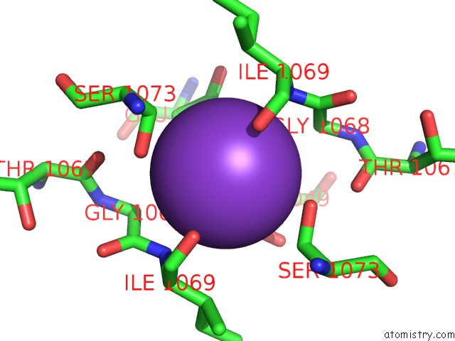
Mono view
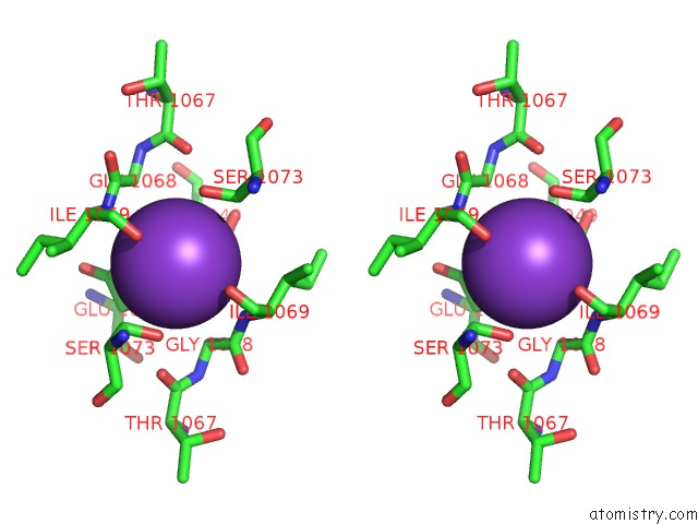
Stereo pair view
Mono view
Stereo pair view
A full contact list of Potassium with other atoms in the K binding
site number 1 of X-Ray Structure of the Uridine Phosphorylase From Salmonella Typhimurium in Complex with 5-Fluorouracil at 2.15 A Resolution within 5.0Å range:
|
Potassium binding site 2 out of 3 in 4e1v
Go back to
Potassium binding site 2 out
of 3 in the X-Ray Structure of the Uridine Phosphorylase From Salmonella Typhimurium in Complex with 5-Fluorouracil at 2.15 A Resolution
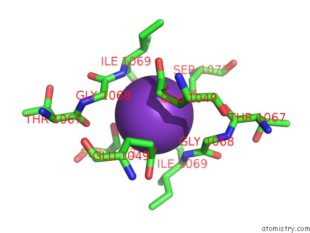
Mono view
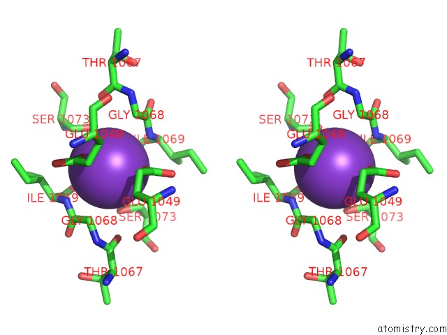
Stereo pair view
Mono view
Stereo pair view
A full contact list of Potassium with other atoms in the K binding
site number 2 of X-Ray Structure of the Uridine Phosphorylase From Salmonella Typhimurium in Complex with 5-Fluorouracil at 2.15 A Resolution within 5.0Å range:
|
Potassium binding site 3 out of 3 in 4e1v
Go back to
Potassium binding site 3 out
of 3 in the X-Ray Structure of the Uridine Phosphorylase From Salmonella Typhimurium in Complex with 5-Fluorouracil at 2.15 A Resolution
Mono view
Stereo pair view
Mono view
Stereo pair view
A full contact list of Potassium with other atoms in the K binding
site number 3 of X-Ray Structure of the Uridine Phosphorylase From Salmonella Typhimurium in Complex with 5-Fluorouracil at 2.15 A Resolution within 5.0Å range:
|
Reference:
A.A.Lashkov,
S.E.Sotnichenko,
I.I.Prokofiev,
A.G.Gabdulkhakov,
I.I.Agapov,
A.A.Shtil,
C.Betzel,
A.S.Mironov,
A.M.Mikhailov.
X-Ray Structure of Salmonella Typhimurium Uridine Phosphorylase Complexed with 5-Fluorouracil and Molecular Modelling of the Complex of 5-Fluorouracil with Uridine Phosphorylase From Vibrio Cholerae. Acta Crystallogr.,Sect.D V. 68 968 2012.
ISSN: ISSN 0907-4449
PubMed: 22868762
DOI: 10.1107/S090744491201815X
Page generated: Sat Aug 9 06:40:28 2025
ISSN: ISSN 0907-4449
PubMed: 22868762
DOI: 10.1107/S090744491201815X
Last articles
K in 6JJGK in 6JJF
K in 6JJE
K in 6J6T
K in 6JIG
K in 6ISW
K in 6IP3
K in 6IP7
K in 6IAX
K in 6I9D