Potassium »
PDB 6i4i-6lab »
6isw »
Potassium in PDB 6isw: Structure of Human Telomeric Dna with 5-Selenophene-Modified Deoxyuridine at Residue 12
Protein crystallography data
The structure of Structure of Human Telomeric Dna with 5-Selenophene-Modified Deoxyuridine at Residue 12, PDB code: 6isw
was solved by
K.Saikrishnan,
A.Nuthanakanti,
S.G.Srivatsan,
I.Ahmad,
with X-Ray Crystallography technique. A brief refinement statistics is given in the table below:
| Resolution Low / High (Å) | 42.28 / 2.30 |
| Space group | P 21 2 21 |
| Cell size a, b, c (Å), α, β, γ (°) | 35.204, 42.280, 49.924, 90.00, 90.00, 90.00 |
| R / Rfree (%) | 25.2 / 28.3 |
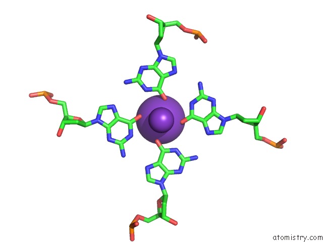
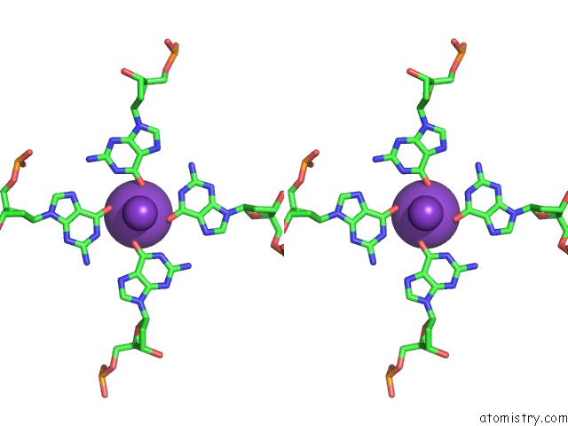
Potassium Binding Sites:
The binding sites of Potassium atom in the Structure of Human Telomeric Dna with 5-Selenophene-Modified Deoxyuridine at Residue 12
(pdb code 6isw). This binding sites where shown within
5.0 Angstroms radius around Potassium atom.
In total 3 binding sites of Potassium where determined in the Structure of Human Telomeric Dna with 5-Selenophene-Modified Deoxyuridine at Residue 12, PDB code: 6isw:
Jump to Potassium binding site number: 1; 2; 3;
In total 3 binding sites of Potassium where determined in the Structure of Human Telomeric Dna with 5-Selenophene-Modified Deoxyuridine at Residue 12, PDB code: 6isw:
Jump to Potassium binding site number: 1; 2; 3;
Potassium binding site 1 out of 3 in 6isw
Go back to
Potassium binding site 1 out
of 3 in the Structure of Human Telomeric Dna with 5-Selenophene-Modified Deoxyuridine at Residue 12
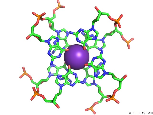
Mono view
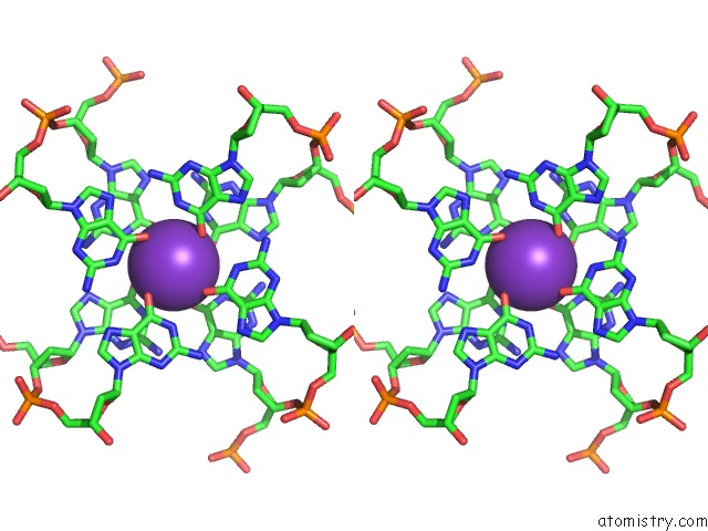
Stereo pair view
Mono view
Stereo pair view
A full contact list of Potassium with other atoms in the K binding
site number 1 of Structure of Human Telomeric Dna with 5-Selenophene-Modified Deoxyuridine at Residue 12 within 5.0Å range:
|
Potassium binding site 2 out of 3 in 6isw
Go back to
Potassium binding site 2 out
of 3 in the Structure of Human Telomeric Dna with 5-Selenophene-Modified Deoxyuridine at Residue 12
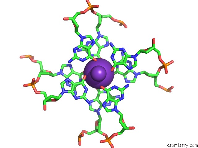
Mono view
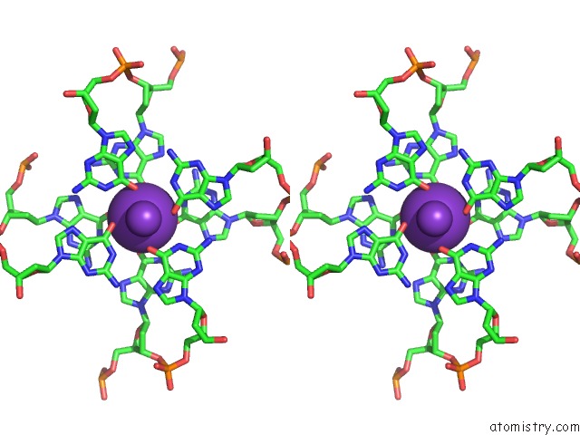
Stereo pair view
Mono view
Stereo pair view
A full contact list of Potassium with other atoms in the K binding
site number 2 of Structure of Human Telomeric Dna with 5-Selenophene-Modified Deoxyuridine at Residue 12 within 5.0Å range:
|
Potassium binding site 3 out of 3 in 6isw
Go back to
Potassium binding site 3 out
of 3 in the Structure of Human Telomeric Dna with 5-Selenophene-Modified Deoxyuridine at Residue 12
Mono view
Stereo pair view
Mono view
Stereo pair view
A full contact list of Potassium with other atoms in the K binding
site number 3 of Structure of Human Telomeric Dna with 5-Selenophene-Modified Deoxyuridine at Residue 12 within 5.0Å range:
|
Reference:
A.Nuthanakanti,
I.Ahmed,
S.Y.Khatik,
K.Saikrishnan,
S.G.Srivatsan.
Probing G-Quadruplex Topologies and Recognition Concurrently in Real Time and 3D Using A Dual-App Nucleoside Probe. Nucleic Acids Res. V. 47 6059 2019.
ISSN: ESSN 1362-4962
PubMed: 31106340
DOI: 10.1093/NAR/GKZ419
Page generated: Sat Aug 9 11:25:28 2025
ISSN: ESSN 1362-4962
PubMed: 31106340
DOI: 10.1093/NAR/GKZ419
Last articles
Mo in 2VPZMo in 2VPY
Mo in 2VPX
Mo in 2VPW
Mo in 2MIN
Mo in 2V45
Mo in 2V3V
Mo in 2NYA
Mo in 2R8U
Mo in 2ONR