Potassium »
PDB 4bga-4cn0 »
4bz6 »
Potassium in PDB 4bz6: Crystal Structure of Schistosoma Mansoni HDAC8 Complexed with Saha
Protein crystallography data
The structure of Crystal Structure of Schistosoma Mansoni HDAC8 Complexed with Saha, PDB code: 4bz6
was solved by
M.Marek,
C.Romier,
with X-Ray Crystallography technique. A brief refinement statistics is given in the table below:
| Resolution Low / High (Å) | 34.74 / 2.00 |
| Space group | P 1 |
| Cell size a, b, c (Å), α, β, γ (°) | 70.640, 70.640, 97.990, 77.88, 75.48, 85.69 |
| R / Rfree (%) | 16.33 / 19.1 |
Other elements in 4bz6:
The structure of Crystal Structure of Schistosoma Mansoni HDAC8 Complexed with Saha also contains other interesting chemical elements:
| Zinc | (Zn) | 4 atoms |
Potassium Binding Sites:
The binding sites of Potassium atom in the Crystal Structure of Schistosoma Mansoni HDAC8 Complexed with Saha
(pdb code 4bz6). This binding sites where shown within
5.0 Angstroms radius around Potassium atom.
In total 8 binding sites of Potassium where determined in the Crystal Structure of Schistosoma Mansoni HDAC8 Complexed with Saha, PDB code: 4bz6:
Jump to Potassium binding site number: 1; 2; 3; 4; 5; 6; 7; 8;
In total 8 binding sites of Potassium where determined in the Crystal Structure of Schistosoma Mansoni HDAC8 Complexed with Saha, PDB code: 4bz6:
Jump to Potassium binding site number: 1; 2; 3; 4; 5; 6; 7; 8;
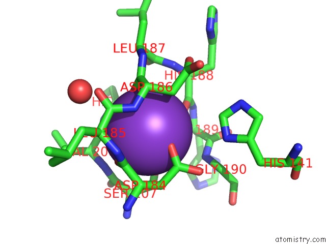
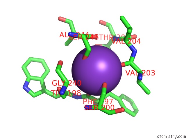
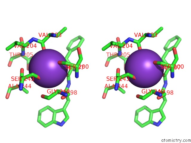
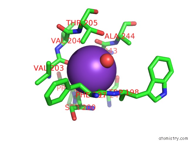
Potassium binding site 1 out of 8 in 4bz6
Go back to
Potassium binding site 1 out
of 8 in the Crystal Structure of Schistosoma Mansoni HDAC8 Complexed with Saha
Mono view
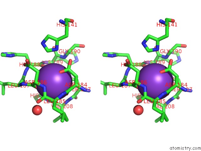
Stereo pair view
Mono view
Stereo pair view
A full contact list of Potassium with other atoms in the K binding
site number 1 of Crystal Structure of Schistosoma Mansoni HDAC8 Complexed with Saha within 5.0Å range:
|
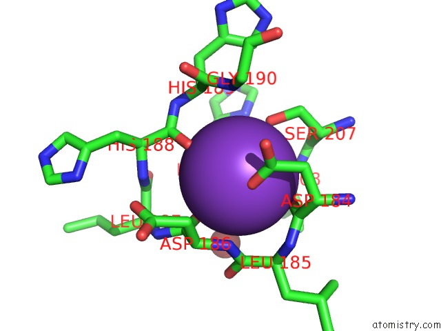
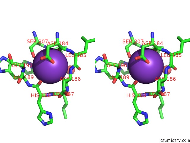
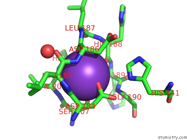
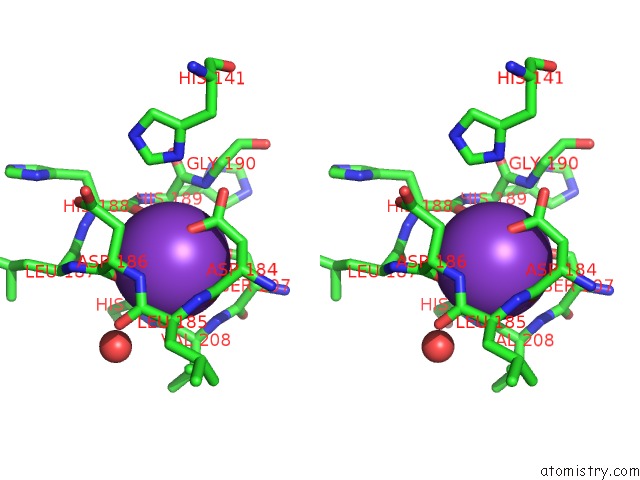
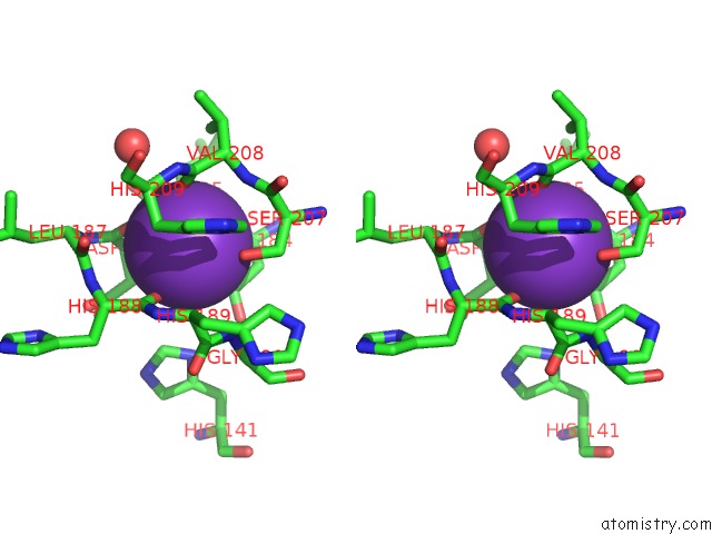
Potassium binding site 2 out of 8 in 4bz6
Go back to
Potassium binding site 2 out
of 8 in the Crystal Structure of Schistosoma Mansoni HDAC8 Complexed with Saha

Mono view

Stereo pair view
Mono view
Stereo pair view
A full contact list of Potassium with other atoms in the K binding
site number 2 of Crystal Structure of Schistosoma Mansoni HDAC8 Complexed with Saha within 5.0Å range:
|
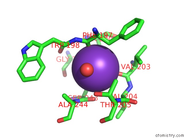
Potassium binding site 3 out of 8 in 4bz6
Go back to
Potassium binding site 3 out
of 8 in the Crystal Structure of Schistosoma Mansoni HDAC8 Complexed with Saha
Mono view
Stereo pair view
Mono view
Stereo pair view
A full contact list of Potassium with other atoms in the K binding
site number 3 of Crystal Structure of Schistosoma Mansoni HDAC8 Complexed with Saha within 5.0Å range:
|
Potassium binding site 4 out of 8 in 4bz6
Go back to
Potassium binding site 4 out
of 8 in the Crystal Structure of Schistosoma Mansoni HDAC8 Complexed with Saha
Mono view
Stereo pair view
Mono view
Stereo pair view
A full contact list of Potassium with other atoms in the K binding
site number 4 of Crystal Structure of Schistosoma Mansoni HDAC8 Complexed with Saha within 5.0Å range:
|
Potassium binding site 5 out of 8 in 4bz6
Go back to
Potassium binding site 5 out
of 8 in the Crystal Structure of Schistosoma Mansoni HDAC8 Complexed with Saha
Mono view
Stereo pair view
Mono view
Stereo pair view
A full contact list of Potassium with other atoms in the K binding
site number 5 of Crystal Structure of Schistosoma Mansoni HDAC8 Complexed with Saha within 5.0Å range:
|
Potassium binding site 6 out of 8 in 4bz6
Go back to
Potassium binding site 6 out
of 8 in the Crystal Structure of Schistosoma Mansoni HDAC8 Complexed with Saha
Mono view

Stereo pair view
Mono view
Stereo pair view
A full contact list of Potassium with other atoms in the K binding
site number 6 of Crystal Structure of Schistosoma Mansoni HDAC8 Complexed with Saha within 5.0Å range:
|
Potassium binding site 7 out of 8 in 4bz6
Go back to
Potassium binding site 7 out
of 8 in the Crystal Structure of Schistosoma Mansoni HDAC8 Complexed with Saha

Mono view
Stereo pair view
Mono view
Stereo pair view
A full contact list of Potassium with other atoms in the K binding
site number 7 of Crystal Structure of Schistosoma Mansoni HDAC8 Complexed with Saha within 5.0Å range:
|
Potassium binding site 8 out of 8 in 4bz6
Go back to
Potassium binding site 8 out
of 8 in the Crystal Structure of Schistosoma Mansoni HDAC8 Complexed with Saha
Mono view

Stereo pair view
Mono view
Stereo pair view
A full contact list of Potassium with other atoms in the K binding
site number 8 of Crystal Structure of Schistosoma Mansoni HDAC8 Complexed with Saha within 5.0Å range:
|
Reference:
M.Marek,
S.Kannan,
A.Hauser,
M.Moraes Mourao,
S.Caby,
V.Cura,
D.A.Stolfa,
K.Schmidtkunz,
J.Lancelot,
L.Andrade,
J.Renaud,
G.Oliveira,
W.Sippl,
M.Jung,
J.Cavarelli,
R.J.Pierce,
C.Romier.
Structural Basis For the Inhibition of Histone Deacetylase 8 (HDAC8), A Key Epigenetic Player in the Blood Fluke Schistosoma Mansoni. Plos Pathog. V. 9 03645 2013.
ISSN: ISSN 1553-7366
PubMed: 24086136
DOI: 10.1371/JOURNAL.PPAT.1003645
Page generated: Mon Aug 12 10:16:04 2024
ISSN: ISSN 1553-7366
PubMed: 24086136
DOI: 10.1371/JOURNAL.PPAT.1003645
Last articles
Zn in 9J0NZn in 9J0O
Zn in 9J0P
Zn in 9FJX
Zn in 9EKB
Zn in 9C0F
Zn in 9CAH
Zn in 9CH0
Zn in 9CH3
Zn in 9CH1