Potassium »
PDB 4bga-4cn0 »
4bva »
Potassium in PDB 4bva: Crystal Structure of the Nadph-T3 Form of Mouse Mu-Crystallin.
Enzymatic activity of Crystal Structure of the Nadph-T3 Form of Mouse Mu-Crystallin.
All present enzymatic activity of Crystal Structure of the Nadph-T3 Form of Mouse Mu-Crystallin.:
1.5.1.25;
1.5.1.25;
Protein crystallography data
The structure of Crystal Structure of the Nadph-T3 Form of Mouse Mu-Crystallin., PDB code: 4bva
was solved by
F.Borel,
I.Hachi,
A.Palencia,
M.C.Gaillard,
J.L.Ferrer,
with X-Ray Crystallography technique. A brief refinement statistics is given in the table below:
| Resolution Low / High (Å) | 42.68 / 1.75 |
| Space group | P 1 21 1 |
| Cell size a, b, c (Å), α, β, γ (°) | 45.240, 97.140, 75.670, 90.00, 104.90, 90.00 |
| R / Rfree (%) | 14.624 / 19.224 |
Other elements in 4bva:
The structure of Crystal Structure of the Nadph-T3 Form of Mouse Mu-Crystallin. also contains other interesting chemical elements:
| Iodine | (I) | 12 atoms |
Potassium Binding Sites:
The binding sites of Potassium atom in the Crystal Structure of the Nadph-T3 Form of Mouse Mu-Crystallin.
(pdb code 4bva). This binding sites where shown within
5.0 Angstroms radius around Potassium atom.
In total 2 binding sites of Potassium where determined in the Crystal Structure of the Nadph-T3 Form of Mouse Mu-Crystallin., PDB code: 4bva:
Jump to Potassium binding site number: 1; 2;
In total 2 binding sites of Potassium where determined in the Crystal Structure of the Nadph-T3 Form of Mouse Mu-Crystallin., PDB code: 4bva:
Jump to Potassium binding site number: 1; 2;
Potassium binding site 1 out of 2 in 4bva
Go back to
Potassium binding site 1 out
of 2 in the Crystal Structure of the Nadph-T3 Form of Mouse Mu-Crystallin.
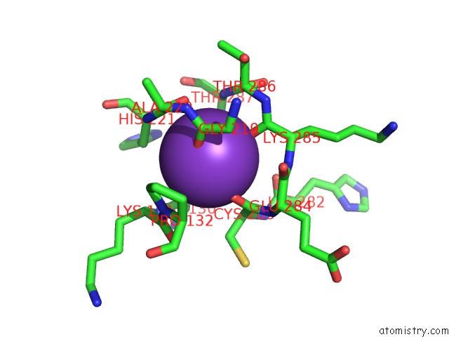
Mono view
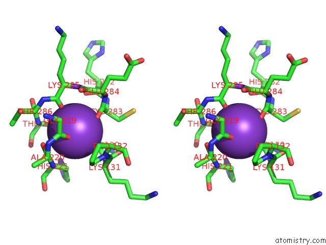
Stereo pair view
Mono view
Stereo pair view
A full contact list of Potassium with other atoms in the K binding
site number 1 of Crystal Structure of the Nadph-T3 Form of Mouse Mu-Crystallin. within 5.0Å range:
|
Potassium binding site 2 out of 2 in 4bva
Go back to
Potassium binding site 2 out
of 2 in the Crystal Structure of the Nadph-T3 Form of Mouse Mu-Crystallin.
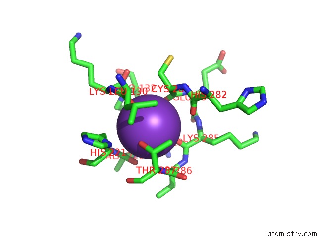
Mono view
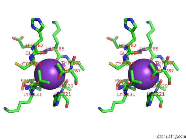
Stereo pair view
Mono view
Stereo pair view
A full contact list of Potassium with other atoms in the K binding
site number 2 of Crystal Structure of the Nadph-T3 Form of Mouse Mu-Crystallin. within 5.0Å range:
|
Reference:
F.Borel,
I.Hachi,
A.Palencia,
M.C.Gaillard,
J.L.Ferrer.
Crystal Structure of Mouse Mu-Crystallin Complexed with Nadph and the T3 Thyroid Hormone Febs J. V. 281 1598 2014.
ISSN: ISSN 1742-464X
PubMed: 24467707
DOI: 10.1111/FEBS.12726
Page generated: Mon Aug 12 10:13:09 2024
ISSN: ISSN 1742-464X
PubMed: 24467707
DOI: 10.1111/FEBS.12726
Last articles
Zn in 9J0NZn in 9J0O
Zn in 9J0P
Zn in 9FJX
Zn in 9EKB
Zn in 9C0F
Zn in 9CAH
Zn in 9CH0
Zn in 9CH3
Zn in 9CH1