Potassium »
PDB 4bga-4cn0 »
4bi3 »
Potassium in PDB 4bi3: Structure and Function of Amidase Toxin - Antitoxin Combinations Associated with the Type VI Secretion System of Serratia Marcescens.
Protein crystallography data
The structure of Structure and Function of Amidase Toxin - Antitoxin Combinations Associated with the Type VI Secretion System of Serratia Marcescens., PDB code: 4bi3
was solved by
V.Srikannathasan,
G.English,
N.K.Bui,
K.Trunk,
P.E.F.O.Rourke,
V.A.Rao,
W.Vollmer,
S.J.Coulthurst,
W.N.Hunter,
with X-Ray Crystallography technique. A brief refinement statistics is given in the table below:
| Resolution Low / High (Å) | 54.23 / 1.85 |
| Space group | P 21 21 21 |
| Cell size a, b, c (Å), α, β, γ (°) | 56.828, 65.260, 97.504, 90.00, 90.00, 90.00 |
| R / Rfree (%) | 20.732 / 23.685 |
Potassium Binding Sites:
The binding sites of Potassium atom in the Structure and Function of Amidase Toxin - Antitoxin Combinations Associated with the Type VI Secretion System of Serratia Marcescens.
(pdb code 4bi3). This binding sites where shown within
5.0 Angstroms radius around Potassium atom.
In total 3 binding sites of Potassium where determined in the Structure and Function of Amidase Toxin - Antitoxin Combinations Associated with the Type VI Secretion System of Serratia Marcescens., PDB code: 4bi3:
Jump to Potassium binding site number: 1; 2; 3;
In total 3 binding sites of Potassium where determined in the Structure and Function of Amidase Toxin - Antitoxin Combinations Associated with the Type VI Secretion System of Serratia Marcescens., PDB code: 4bi3:
Jump to Potassium binding site number: 1; 2; 3;
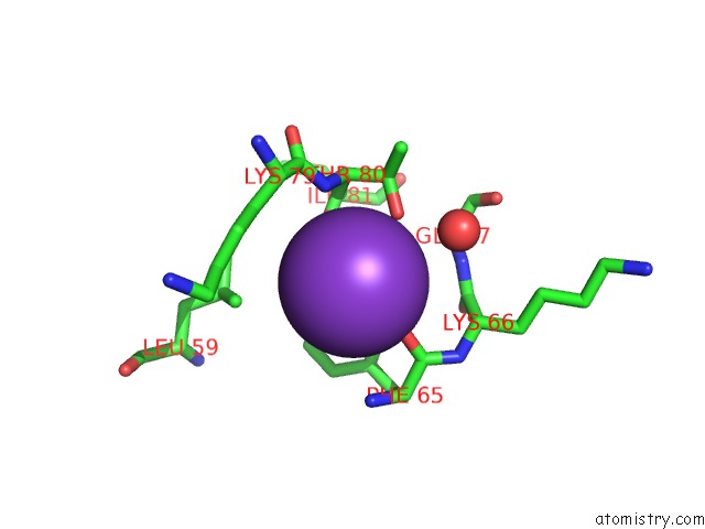
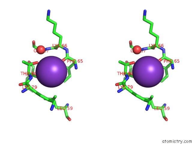
Potassium binding site 1 out of 3 in 4bi3
Go back to
Potassium binding site 1 out
of 3 in the Structure and Function of Amidase Toxin - Antitoxin Combinations Associated with the Type VI Secretion System of Serratia Marcescens.
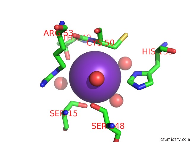
Mono view
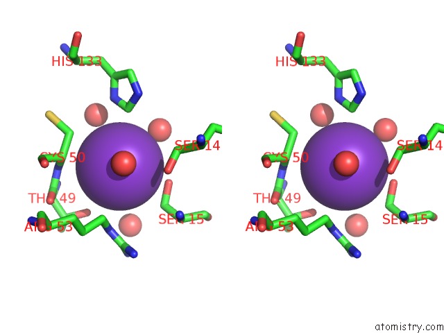
Stereo pair view
Mono view
Stereo pair view
A full contact list of Potassium with other atoms in the K binding
site number 1 of Structure and Function of Amidase Toxin - Antitoxin Combinations Associated with the Type VI Secretion System of Serratia Marcescens. within 5.0Å range:
|
Potassium binding site 2 out of 3 in 4bi3
Go back to
Potassium binding site 2 out
of 3 in the Structure and Function of Amidase Toxin - Antitoxin Combinations Associated with the Type VI Secretion System of Serratia Marcescens.
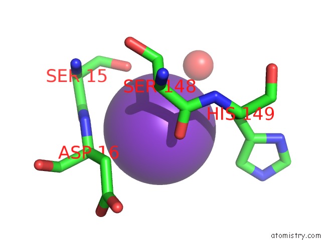
Mono view
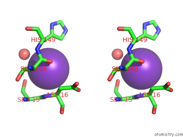
Stereo pair view
Mono view
Stereo pair view
A full contact list of Potassium with other atoms in the K binding
site number 2 of Structure and Function of Amidase Toxin - Antitoxin Combinations Associated with the Type VI Secretion System of Serratia Marcescens. within 5.0Å range:
|
Potassium binding site 3 out of 3 in 4bi3
Go back to
Potassium binding site 3 out
of 3 in the Structure and Function of Amidase Toxin - Antitoxin Combinations Associated with the Type VI Secretion System of Serratia Marcescens.
Mono view
Stereo pair view
Mono view
Stereo pair view
A full contact list of Potassium with other atoms in the K binding
site number 3 of Structure and Function of Amidase Toxin - Antitoxin Combinations Associated with the Type VI Secretion System of Serratia Marcescens. within 5.0Å range:
|
Reference:
V.Srikannathasan,
G.English,
N.K.Bui,
K.Trunk,
P.E.F.O.Rourke,
V.A.Rao,
W.Vollmer,
S.J.Coulthurst,
W.N.Hunter.
Structural Basis For Type VI Secreted Peptidoglycan Dl-Endopeptidase Function, Specificity and Neutralization in Serratia Marcescens Acta Crystallogr.,Sect.D V. 69 2468 2013.
ISSN: ISSN 0907-4449
PubMed: 24311588
DOI: 10.1107/S0907444913022725
Page generated: Mon Aug 12 10:11:55 2024
ISSN: ISSN 0907-4449
PubMed: 24311588
DOI: 10.1107/S0907444913022725
Last articles
Zn in 9J0NZn in 9J0O
Zn in 9J0P
Zn in 9FJX
Zn in 9EKB
Zn in 9C0F
Zn in 9CAH
Zn in 9CH0
Zn in 9CH3
Zn in 9CH1