Potassium »
PDB 1yjn-2aaq »
2a1o »
Potassium in PDB 2a1o: Crystal Structure of Ferrous Dioxygen Complex of T252A Cytochrome P450CAM
Enzymatic activity of Crystal Structure of Ferrous Dioxygen Complex of T252A Cytochrome P450CAM
All present enzymatic activity of Crystal Structure of Ferrous Dioxygen Complex of T252A Cytochrome P450CAM:
1.14.15.1;
1.14.15.1;
Protein crystallography data
The structure of Crystal Structure of Ferrous Dioxygen Complex of T252A Cytochrome P450CAM, PDB code: 2a1o
was solved by
S.Nagano,
T.L.Poulos,
with X-Ray Crystallography technique. A brief refinement statistics is given in the table below:
| Resolution Low / High (Å) | 41.07 / 1.55 |
| Space group | P 1 21 1 |
| Cell size a, b, c (Å), α, β, γ (°) | 67.260, 62.180, 95.220, 90.00, 90.53, 90.00 |
| R / Rfree (%) | 19.7 / 21.9 |
Other elements in 2a1o:
The structure of Crystal Structure of Ferrous Dioxygen Complex of T252A Cytochrome P450CAM also contains other interesting chemical elements:
| Iron | (Fe) | 2 atoms |
Potassium Binding Sites:
The binding sites of Potassium atom in the Crystal Structure of Ferrous Dioxygen Complex of T252A Cytochrome P450CAM
(pdb code 2a1o). This binding sites where shown within
5.0 Angstroms radius around Potassium atom.
In total 3 binding sites of Potassium where determined in the Crystal Structure of Ferrous Dioxygen Complex of T252A Cytochrome P450CAM, PDB code: 2a1o:
Jump to Potassium binding site number: 1; 2; 3;
In total 3 binding sites of Potassium where determined in the Crystal Structure of Ferrous Dioxygen Complex of T252A Cytochrome P450CAM, PDB code: 2a1o:
Jump to Potassium binding site number: 1; 2; 3;
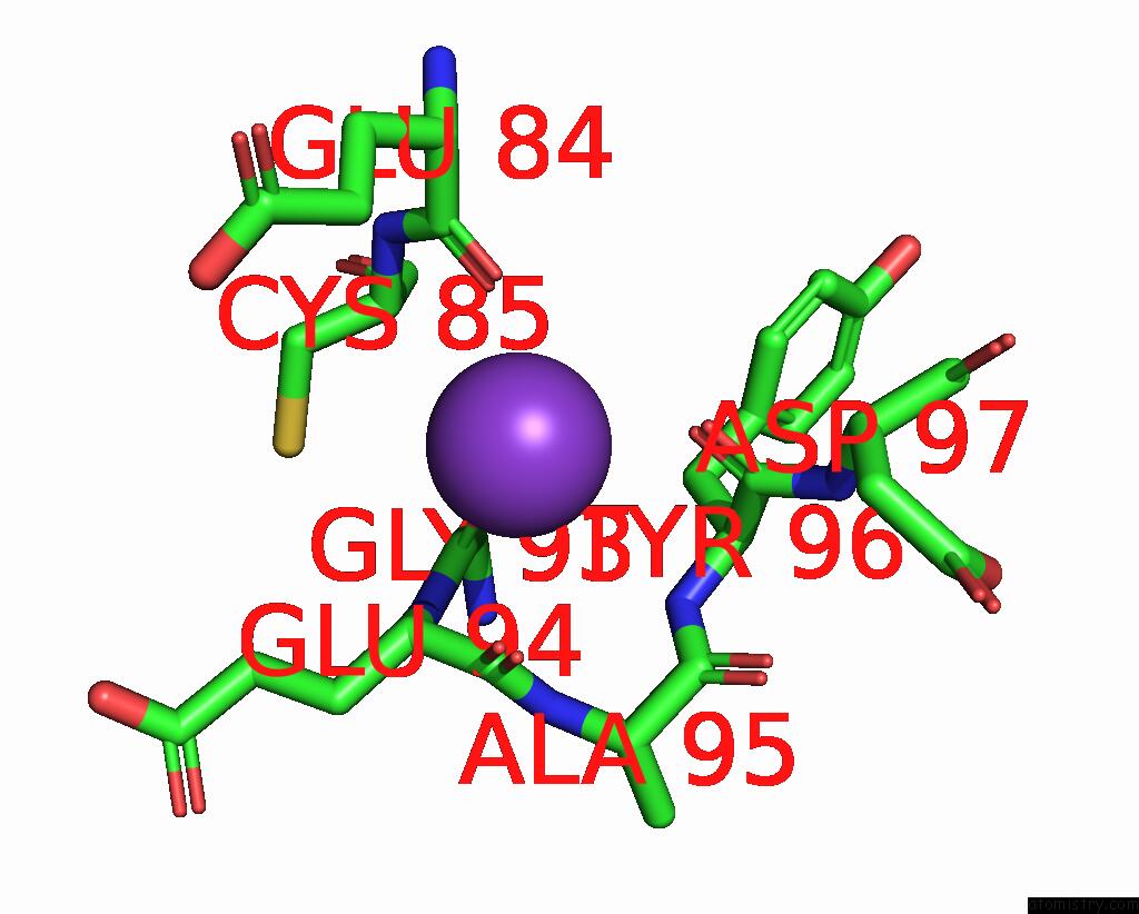
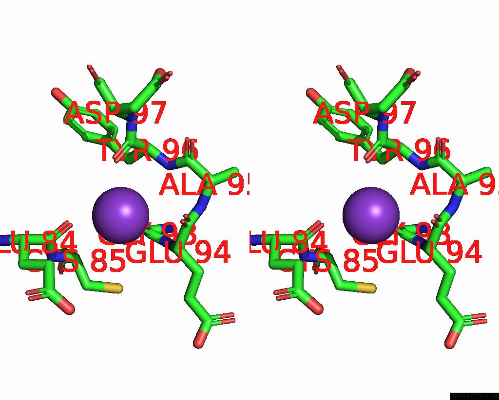
Potassium binding site 1 out of 3 in 2a1o
Go back to
Potassium binding site 1 out
of 3 in the Crystal Structure of Ferrous Dioxygen Complex of T252A Cytochrome P450CAM
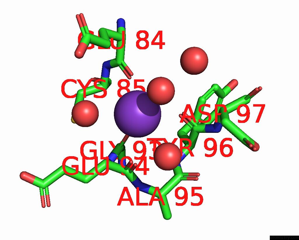
Mono view
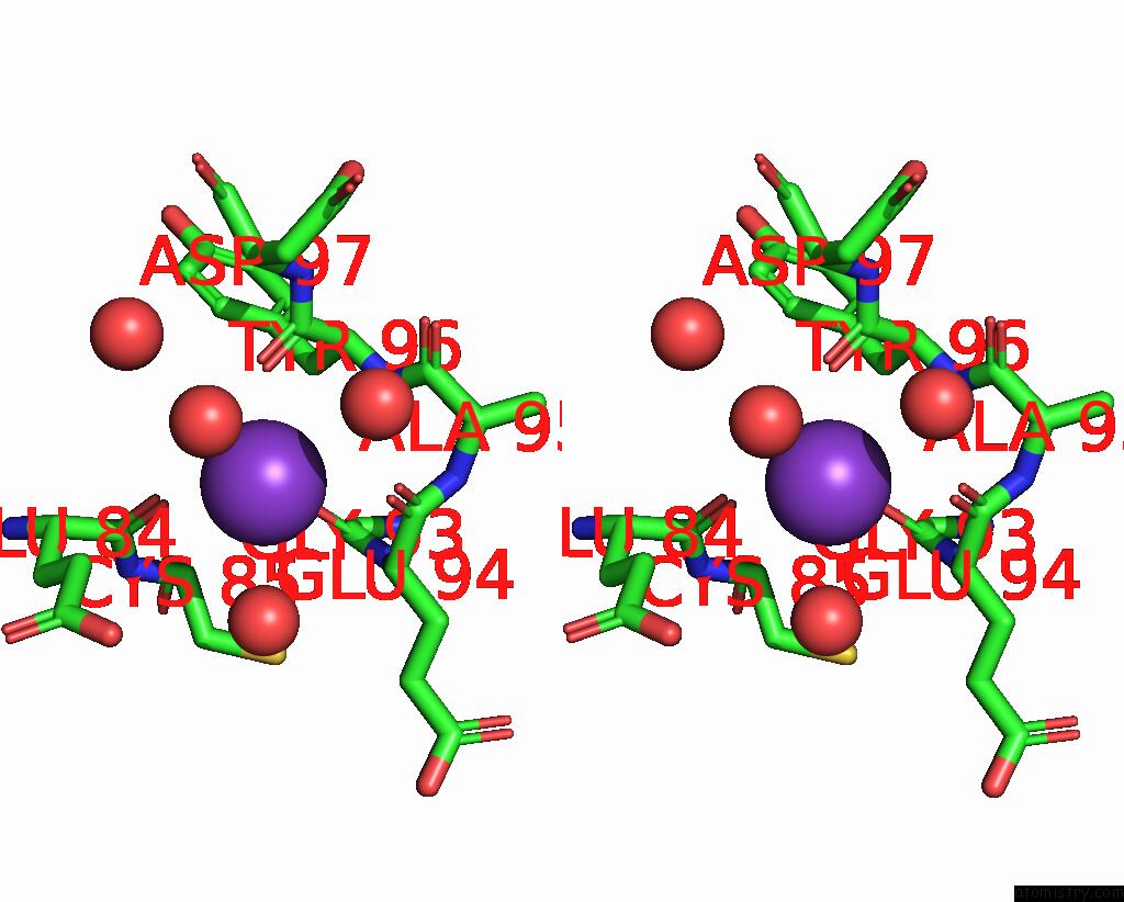
Stereo pair view
Mono view
Stereo pair view
A full contact list of Potassium with other atoms in the K binding
site number 1 of Crystal Structure of Ferrous Dioxygen Complex of T252A Cytochrome P450CAM within 5.0Å range:
|
Potassium binding site 2 out of 3 in 2a1o
Go back to
Potassium binding site 2 out
of 3 in the Crystal Structure of Ferrous Dioxygen Complex of T252A Cytochrome P450CAM
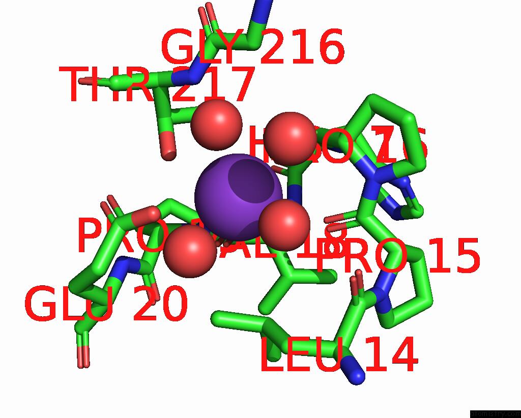
Mono view
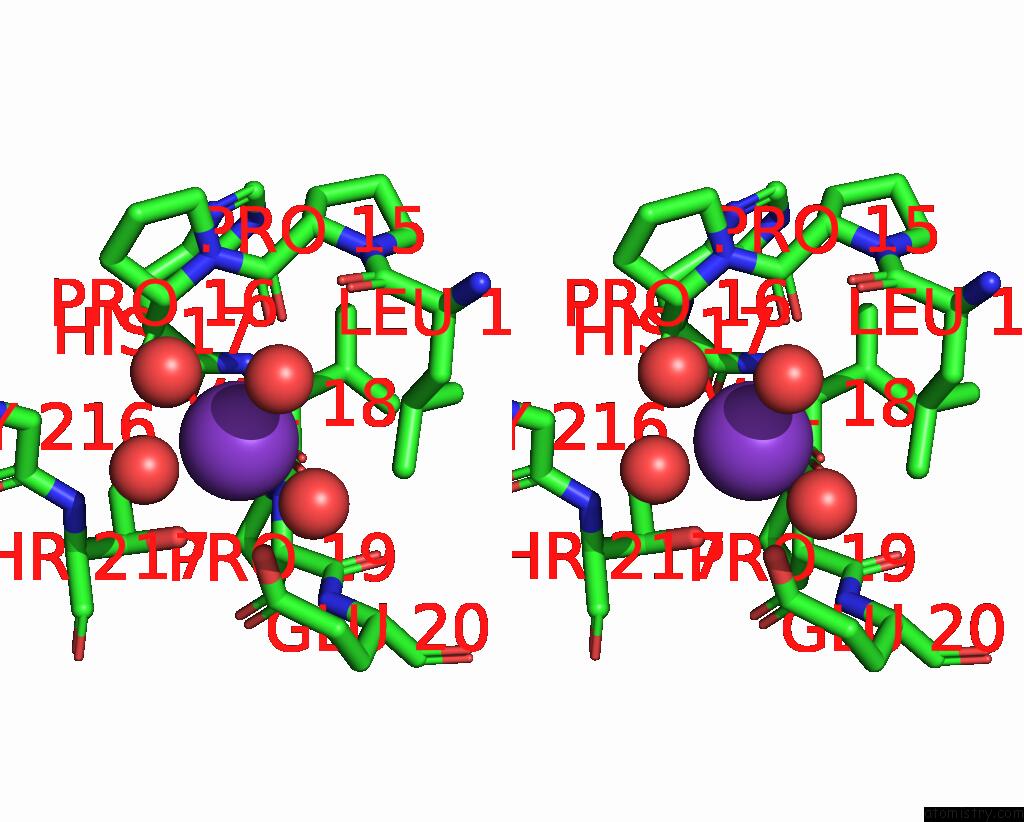
Stereo pair view
Mono view
Stereo pair view
A full contact list of Potassium with other atoms in the K binding
site number 2 of Crystal Structure of Ferrous Dioxygen Complex of T252A Cytochrome P450CAM within 5.0Å range:
|
Potassium binding site 3 out of 3 in 2a1o
Go back to
Potassium binding site 3 out
of 3 in the Crystal Structure of Ferrous Dioxygen Complex of T252A Cytochrome P450CAM
Mono view
Stereo pair view
Mono view
Stereo pair view
A full contact list of Potassium with other atoms in the K binding
site number 3 of Crystal Structure of Ferrous Dioxygen Complex of T252A Cytochrome P450CAM within 5.0Å range:
|
Reference:
S.Nagano,
T.L.Poulos.
Crystallographic Study on the Dioxygen Complex of Wild-Type and Mutant Cytochrome P450CAM. Implications For the Dioxygen Activation Mechanism J.Biol.Chem. V. 280 31659 2005.
ISSN: ISSN 0021-9258
PubMed: 15994329
DOI: 10.1074/JBC.M505261200
Page generated: Sat Aug 9 03:04:53 2025
ISSN: ISSN 0021-9258
PubMed: 15994329
DOI: 10.1074/JBC.M505261200
Last articles
K in 7BVZK in 7BQG
K in 7BVY
K in 7BVU
K in 7BT2
K in 7BMZ
K in 7BMU
K in 7BH2
K in 7BIL
K in 7BI1