Potassium »
PDB 7akk-7byn »
7bqg »
Potassium in PDB 7bqg: Complex Structure of SAV1 and Dendrin
Protein crystallography data
The structure of Complex Structure of SAV1 and Dendrin, PDB code: 7bqg
was solved by
Z.Lin,
M.Zhang,
with X-Ray Crystallography technique. A brief refinement statistics is given in the table below:
| Resolution Low / High (Å) | 49.69 / 1.55 |
| Space group | C 2 2 21 |
| Cell size a, b, c (Å), α, β, γ (°) | 38.803, 43.084, 99.385, 90.00, 90.00, 90.00 |
| R / Rfree (%) | 17.8 / 21.7 |
Potassium Binding Sites:
The binding sites of Potassium atom in the Complex Structure of SAV1 and Dendrin
(pdb code 7bqg). This binding sites where shown within
5.0 Angstroms radius around Potassium atom.
In total 3 binding sites of Potassium where determined in the Complex Structure of SAV1 and Dendrin, PDB code: 7bqg:
Jump to Potassium binding site number: 1; 2; 3;
In total 3 binding sites of Potassium where determined in the Complex Structure of SAV1 and Dendrin, PDB code: 7bqg:
Jump to Potassium binding site number: 1; 2; 3;
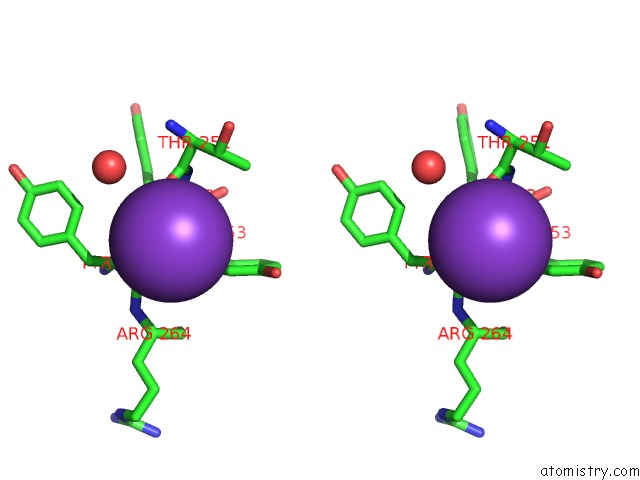
Potassium binding site 1 out of 3 in 7bqg
Go back to
Potassium binding site 1 out
of 3 in the Complex Structure of SAV1 and Dendrin

Mono view
Stereo pair view
Mono view
Stereo pair view
A full contact list of Potassium with other atoms in the K binding
site number 1 of Complex Structure of SAV1 and Dendrin within 5.0Å range:
|
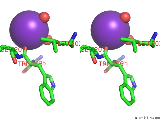
Potassium binding site 2 out of 3 in 7bqg
Go back to
Potassium binding site 2 out
of 3 in the Complex Structure of SAV1 and Dendrin

Mono view
Stereo pair view
Mono view
Stereo pair view
A full contact list of Potassium with other atoms in the K binding
site number 2 of Complex Structure of SAV1 and Dendrin within 5.0Å range:
|
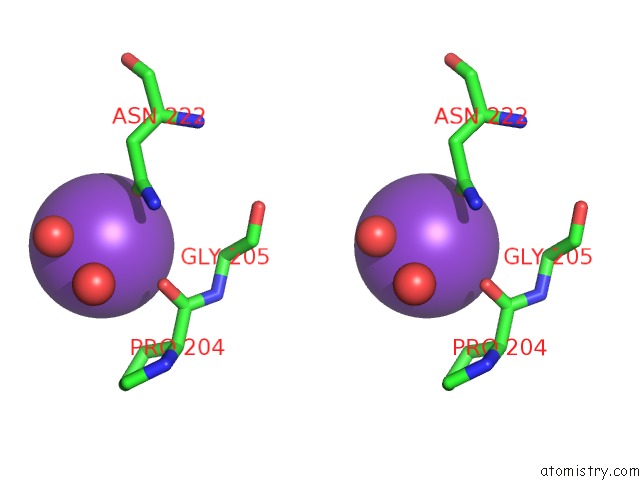
Potassium binding site 3 out of 3 in 7bqg
Go back to
Potassium binding site 3 out
of 3 in the Complex Structure of SAV1 and Dendrin

Mono view
Stereo pair view
Mono view
Stereo pair view
A full contact list of Potassium with other atoms in the K binding
site number 3 of Complex Structure of SAV1 and Dendrin within 5.0Å range:
|
Reference:
Z.Lin,
R.Xie,
K.Guan,
M.Zhang.
A Ww Tandem-Mediated Dimerization Mode of SAV1 Essential For Hippo Signaling. Cell Rep V. 32 08118 2020.
ISSN: ESSN 2211-1247
PubMed: 32905778
DOI: 10.1016/J.CELREP.2020.108118
Page generated: Mon Aug 12 18:45:52 2024
ISSN: ESSN 2211-1247
PubMed: 32905778
DOI: 10.1016/J.CELREP.2020.108118
Last articles
Fe in 4UQHFe in 4UPT
Fe in 4UPS
Fe in 4UPR
Fe in 4UPQ
Fe in 4UPP
Fe in 4UPO
Fe in 4UOB
Fe in 4UNF
Fe in 4UPN