Potassium »
PDB 1k4d-1m40 »
1l1h »
Potassium in PDB 1l1h: Crystal Structure of the Quadruplex Dna-Drug Complex
Protein crystallography data
The structure of Crystal Structure of the Quadruplex Dna-Drug Complex, PDB code: 1l1h
was solved by
S.M.Haider,
G.N.Parkinson,
S.Neidle,
with X-Ray Crystallography technique. A brief refinement statistics is given in the table below:
| Resolution Low / High (Å) | 10.00 / 1.75 |
| Space group | P 21 21 2 |
| Cell size a, b, c (Å), α, β, γ (°) | 55.451, 42.736, 26.926, 90.00, 90.00, 90.00 |
| R / Rfree (%) | 14.4 / 21.9 |
Potassium Binding Sites:
The binding sites of Potassium atom in the Crystal Structure of the Quadruplex Dna-Drug Complex
(pdb code 1l1h). This binding sites where shown within
5.0 Angstroms radius around Potassium atom.
In total 4 binding sites of Potassium where determined in the Crystal Structure of the Quadruplex Dna-Drug Complex, PDB code: 1l1h:
Jump to Potassium binding site number: 1; 2; 3; 4;
In total 4 binding sites of Potassium where determined in the Crystal Structure of the Quadruplex Dna-Drug Complex, PDB code: 1l1h:
Jump to Potassium binding site number: 1; 2; 3; 4;
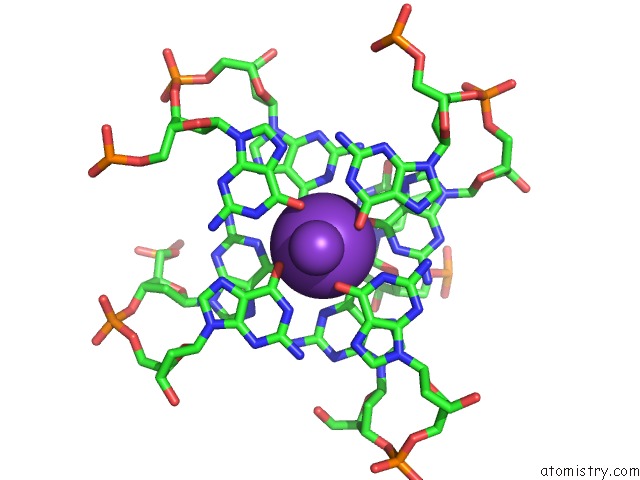
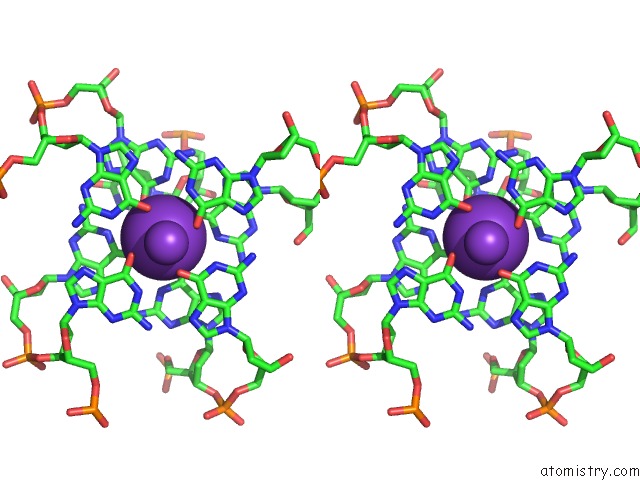
Potassium binding site 1 out of 4 in 1l1h
Go back to
Potassium binding site 1 out
of 4 in the Crystal Structure of the Quadruplex Dna-Drug Complex
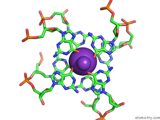
Mono view
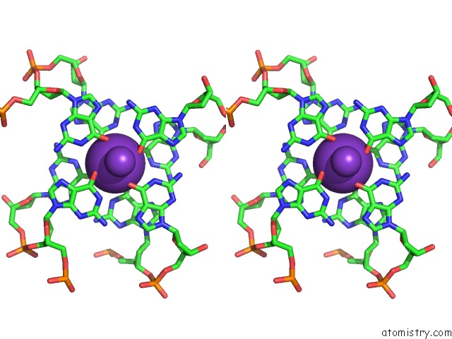
Stereo pair view
Mono view
Stereo pair view
A full contact list of Potassium with other atoms in the K binding
site number 1 of Crystal Structure of the Quadruplex Dna-Drug Complex within 5.0Å range:
|
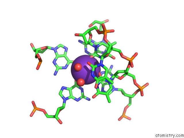
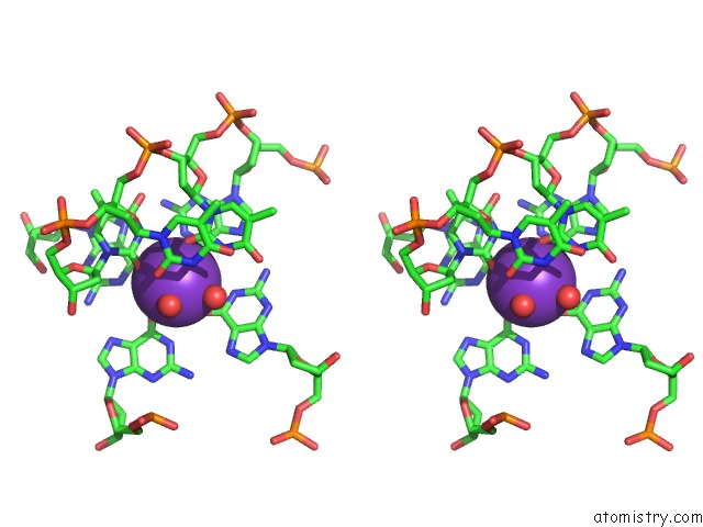
Potassium binding site 2 out of 4 in 1l1h
Go back to
Potassium binding site 2 out
of 4 in the Crystal Structure of the Quadruplex Dna-Drug Complex
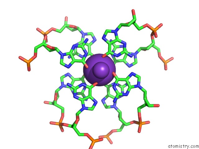
Mono view
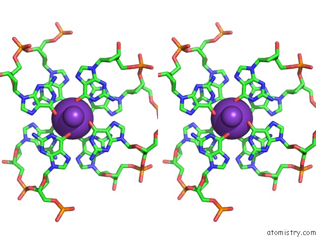
Stereo pair view
Mono view
Stereo pair view
A full contact list of Potassium with other atoms in the K binding
site number 2 of Crystal Structure of the Quadruplex Dna-Drug Complex within 5.0Å range:
|
Potassium binding site 3 out of 4 in 1l1h
Go back to
Potassium binding site 3 out
of 4 in the Crystal Structure of the Quadruplex Dna-Drug Complex
Mono view
Stereo pair view
Mono view
Stereo pair view
A full contact list of Potassium with other atoms in the K binding
site number 3 of Crystal Structure of the Quadruplex Dna-Drug Complex within 5.0Å range:
|
Potassium binding site 4 out of 4 in 1l1h
Go back to
Potassium binding site 4 out
of 4 in the Crystal Structure of the Quadruplex Dna-Drug Complex
Mono view
Stereo pair view
Mono view
Stereo pair view
A full contact list of Potassium with other atoms in the K binding
site number 4 of Crystal Structure of the Quadruplex Dna-Drug Complex within 5.0Å range:
|
Reference:
S.M.Haider,
G.N.Parkinson,
S.Neidle.
Structure of A G-Quadruplex-Ligand Complex J.Mol.Biol. V. 326 117 2003.
ISSN: ISSN 0022-2836
PubMed: 12547195
DOI: 10.1016/S0022-2836(02)01354-2
Page generated: Sat Aug 9 02:09:17 2025
ISSN: ISSN 0022-2836
PubMed: 12547195
DOI: 10.1016/S0022-2836(02)01354-2
Last articles
K in 4BYZK in 4BYG
K in 4BVA
K in 4BGA
K in 4BV8
K in 4BKX
K in 4BGB
K in 4BI3
K in 4BH5
K in 4B3T