Potassium »
PDB 4bga-4cn0 »
4bva »
Potassium in PDB 4bva: Crystal Structure of the Nadph-T3 Form of Mouse Mu-Crystallin.
Enzymatic activity of Crystal Structure of the Nadph-T3 Form of Mouse Mu-Crystallin.
All present enzymatic activity of Crystal Structure of the Nadph-T3 Form of Mouse Mu-Crystallin.:
1.5.1.25;
1.5.1.25;
Protein crystallography data
The structure of Crystal Structure of the Nadph-T3 Form of Mouse Mu-Crystallin., PDB code: 4bva
was solved by
F.Borel,
I.Hachi,
A.Palencia,
M.C.Gaillard,
J.L.Ferrer,
with X-Ray Crystallography technique. A brief refinement statistics is given in the table below:
| Resolution Low / High (Å) | 42.68 / 1.75 |
| Space group | P 1 21 1 |
| Cell size a, b, c (Å), α, β, γ (°) | 45.240, 97.140, 75.670, 90.00, 104.90, 90.00 |
| R / Rfree (%) | 14.624 / 19.224 |
Other elements in 4bva:
The structure of Crystal Structure of the Nadph-T3 Form of Mouse Mu-Crystallin. also contains other interesting chemical elements:
| Iodine | (I) | 12 atoms |
Potassium Binding Sites:
The binding sites of Potassium atom in the Crystal Structure of the Nadph-T3 Form of Mouse Mu-Crystallin.
(pdb code 4bva). This binding sites where shown within
5.0 Angstroms radius around Potassium atom.
In total 2 binding sites of Potassium where determined in the Crystal Structure of the Nadph-T3 Form of Mouse Mu-Crystallin., PDB code: 4bva:
Jump to Potassium binding site number: 1; 2;
In total 2 binding sites of Potassium where determined in the Crystal Structure of the Nadph-T3 Form of Mouse Mu-Crystallin., PDB code: 4bva:
Jump to Potassium binding site number: 1; 2;
Potassium binding site 1 out of 2 in 4bva
Go back to
Potassium binding site 1 out
of 2 in the Crystal Structure of the Nadph-T3 Form of Mouse Mu-Crystallin.
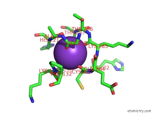
Mono view
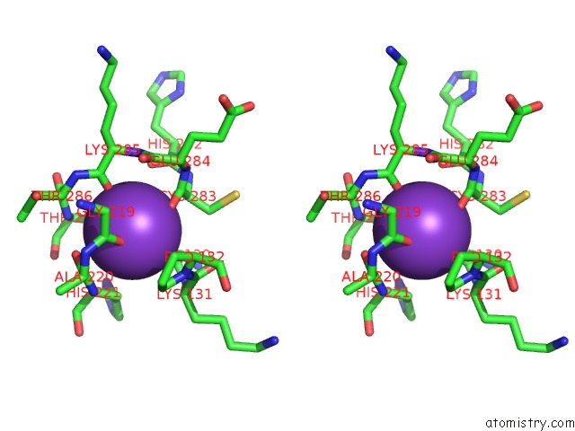
Stereo pair view
Mono view
Stereo pair view
A full contact list of Potassium with other atoms in the K binding
site number 1 of Crystal Structure of the Nadph-T3 Form of Mouse Mu-Crystallin. within 5.0Å range:
|
Potassium binding site 2 out of 2 in 4bva
Go back to
Potassium binding site 2 out
of 2 in the Crystal Structure of the Nadph-T3 Form of Mouse Mu-Crystallin.
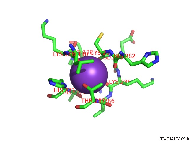
Mono view
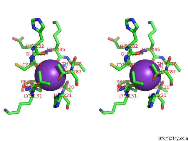
Stereo pair view
Mono view
Stereo pair view
A full contact list of Potassium with other atoms in the K binding
site number 2 of Crystal Structure of the Nadph-T3 Form of Mouse Mu-Crystallin. within 5.0Å range:
|
Reference:
F.Borel,
I.Hachi,
A.Palencia,
M.C.Gaillard,
J.L.Ferrer.
Crystal Structure of Mouse Mu-Crystallin Complexed with Nadph and the T3 Thyroid Hormone Febs J. V. 281 1598 2014.
ISSN: ISSN 1742-464X
PubMed: 24467707
DOI: 10.1111/FEBS.12726
Page generated: Mon Aug 12 10:13:09 2024
ISSN: ISSN 1742-464X
PubMed: 24467707
DOI: 10.1111/FEBS.12726
Last articles
F in 5FRBF in 5FRM
F in 5FR0
F in 5FP0
F in 5FP5
F in 5FHH
F in 5FI4
F in 5FHD
F in 5FI8
F in 5FHE