Potassium »
PDB 1gup-1k4c »
1jdr »
Potassium in PDB 1jdr: Crystal Structure of A Proximal Domain Potassium Binding Variant of Cytochrome C Peroxidase
Enzymatic activity of Crystal Structure of A Proximal Domain Potassium Binding Variant of Cytochrome C Peroxidase
All present enzymatic activity of Crystal Structure of A Proximal Domain Potassium Binding Variant of Cytochrome C Peroxidase:
1.11.1.5;
1.11.1.5;
Protein crystallography data
The structure of Crystal Structure of A Proximal Domain Potassium Binding Variant of Cytochrome C Peroxidase, PDB code: 1jdr
was solved by
C.A.Bonagura,
M.Sundaramoorthy,
B.Bhaskar,
T.L.Poulos,
with X-Ray Crystallography technique. A brief refinement statistics is given in the table below:
| Resolution Low / High (Å) | 10.00 / 1.50 |
| Space group | P 21 21 21 |
| Cell size a, b, c (Å), α, β, γ (°) | 106.717, 75.531, 51.256, 90.00, 90.00, 90.00 |
| R / Rfree (%) | 19.7 / 23.4 |
Other elements in 1jdr:
The structure of Crystal Structure of A Proximal Domain Potassium Binding Variant of Cytochrome C Peroxidase also contains other interesting chemical elements:
| Iron | (Fe) | 1 atom |
Potassium Binding Sites:
The binding sites of Potassium atom in the Crystal Structure of A Proximal Domain Potassium Binding Variant of Cytochrome C Peroxidase
(pdb code 1jdr). This binding sites where shown within
5.0 Angstroms radius around Potassium atom.
In total only one binding site of Potassium was determined in the Crystal Structure of A Proximal Domain Potassium Binding Variant of Cytochrome C Peroxidase, PDB code: 1jdr:
In total only one binding site of Potassium was determined in the Crystal Structure of A Proximal Domain Potassium Binding Variant of Cytochrome C Peroxidase, PDB code: 1jdr:
Potassium binding site 1 out of 1 in 1jdr
Go back to
Potassium binding site 1 out
of 1 in the Crystal Structure of A Proximal Domain Potassium Binding Variant of Cytochrome C Peroxidase
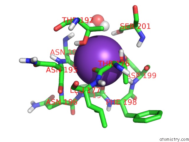
Mono view
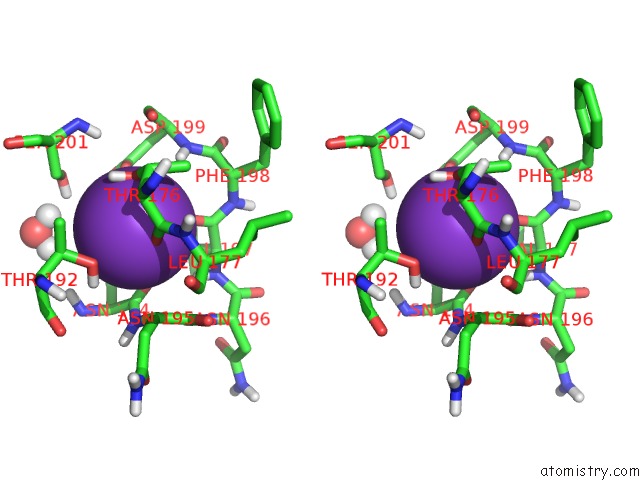
Stereo pair view
Mono view
Stereo pair view
A full contact list of Potassium with other atoms in the K binding
site number 1 of Crystal Structure of A Proximal Domain Potassium Binding Variant of Cytochrome C Peroxidase within 5.0Å range:
|
Reference:
C.A.Bonagura,
M.Sundaramoorthy,
B.Bhaskar,
T.L.Poulos.
The Effects of An Engineered Cation Site on the Structure, Activity, and Epr Properties of Cytochrome C Peroxidase. Biochemistry V. 38 5538 1999.
ISSN: ISSN 0006-2960
PubMed: 10220341
DOI: 10.1021/BI982996K
Page generated: Sat Aug 9 02:01:32 2025
ISSN: ISSN 0006-2960
PubMed: 10220341
DOI: 10.1021/BI982996K
Last articles
K in 5S9LK in 5P9D
K in 5P9C
K in 5P9A
K in 5OX7
K in 5OWO
K in 5OU5
K in 5OSN
K in 5OSX
K in 5OCN