Potassium »
PDB 1d7v-1gmk »
1dz4 »
Potassium in PDB 1dz4: Ferric P450CAM From Pseudomonas Putida
Protein crystallography data
The structure of Ferric P450CAM From Pseudomonas Putida, PDB code: 1dz4
was solved by
I.Schlichting,
J.Berendzen,
K.Chu,
A.M.Stock,
S.A.Maves,
D.E.Benson,
R.M.Sweet,
D.Ringe,
G.A.Petsko,
S.G.Sligar,
with X-Ray Crystallography technique. A brief refinement statistics is given in the table below:
| Resolution Low / High (Å) | 20.00 / 1.60 |
| Space group | P 1 21 1 |
| Cell size a, b, c (Å), α, β, γ (°) | 67.400, 62.700, 95.500, 90.00, 90.65, 90.00 |
| R / Rfree (%) | 18.6 / 23.7 |
Other elements in 1dz4:
The structure of Ferric P450CAM From Pseudomonas Putida also contains other interesting chemical elements:
| Iron | (Fe) | 2 atoms |
Potassium Binding Sites:
The binding sites of Potassium atom in the Ferric P450CAM From Pseudomonas Putida
(pdb code 1dz4). This binding sites where shown within
5.0 Angstroms radius around Potassium atom.
In total 3 binding sites of Potassium where determined in the Ferric P450CAM From Pseudomonas Putida, PDB code: 1dz4:
Jump to Potassium binding site number: 1; 2; 3;
In total 3 binding sites of Potassium where determined in the Ferric P450CAM From Pseudomonas Putida, PDB code: 1dz4:
Jump to Potassium binding site number: 1; 2; 3;
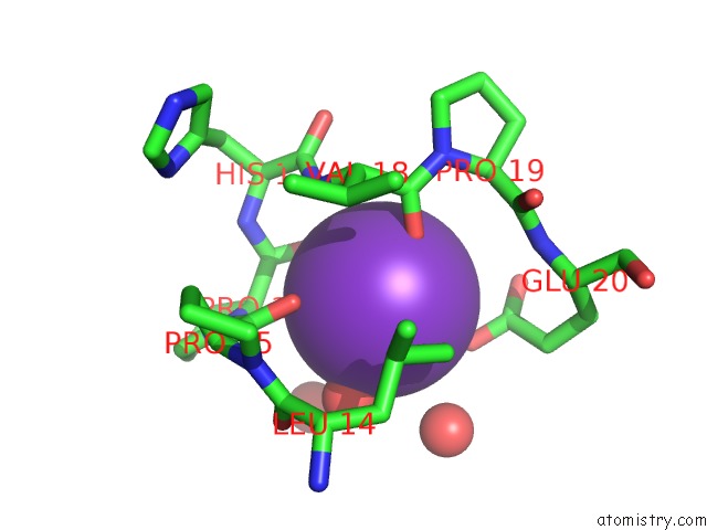
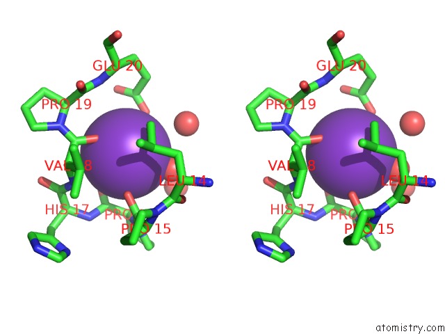
Potassium binding site 1 out of 3 in 1dz4
Go back to
Potassium binding site 1 out
of 3 in the Ferric P450CAM From Pseudomonas Putida
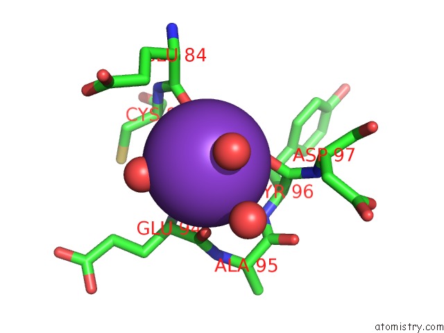
Mono view
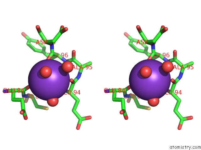
Stereo pair view
Mono view
Stereo pair view
A full contact list of Potassium with other atoms in the K binding
site number 1 of Ferric P450CAM From Pseudomonas Putida within 5.0Å range:
|
Potassium binding site 2 out of 3 in 1dz4
Go back to
Potassium binding site 2 out
of 3 in the Ferric P450CAM From Pseudomonas Putida
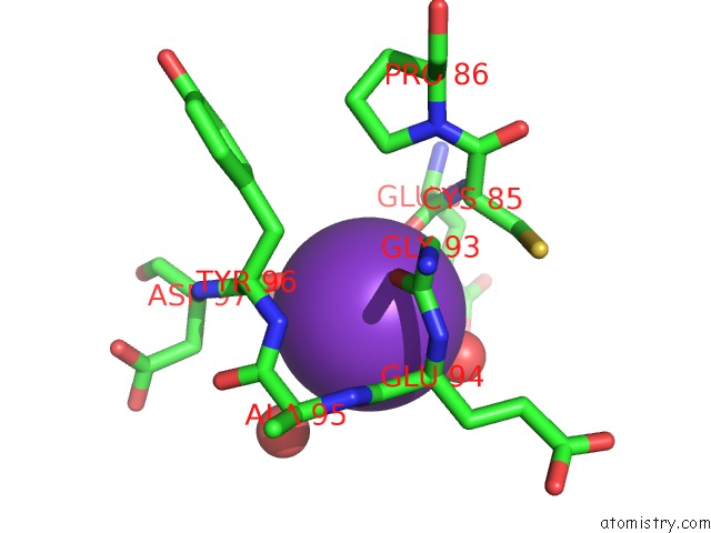
Mono view
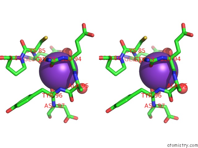
Stereo pair view
Mono view
Stereo pair view
A full contact list of Potassium with other atoms in the K binding
site number 2 of Ferric P450CAM From Pseudomonas Putida within 5.0Å range:
|
Potassium binding site 3 out of 3 in 1dz4
Go back to
Potassium binding site 3 out
of 3 in the Ferric P450CAM From Pseudomonas Putida
Mono view
Stereo pair view
Mono view
Stereo pair view
A full contact list of Potassium with other atoms in the K binding
site number 3 of Ferric P450CAM From Pseudomonas Putida within 5.0Å range:
|
Reference:
I.Schlichting,
J.Berendzen,
K.Chu,
A.M.Stock,
S.A.Maves,
D.E.Benson,
R.M.Sweet,
D.Ringe,
G.A.Petsko,
S.G.Sligar.
The Catalytic Pathway of Cytochrome P450CAM at Atomic Resolution Science V. 287 1615 2000.
ISSN: ISSN 0036-8075
PubMed: 10698731
DOI: 10.1126/SCIENCE.287.5458.1615
Page generated: Sat Aug 9 01:50:29 2025
ISSN: ISSN 0036-8075
PubMed: 10698731
DOI: 10.1126/SCIENCE.287.5458.1615
Last articles
K in 3ZQOK in 3ZNS
K in 3ZNR
K in 3ZED
K in 3WVL
K in 3ZDP
K in 3ZN2
K in 3ZDD
K in 3ZDC
K in 3ZDA