Potassium »
PDB 9jgz-9v0k »
9nec »
Potassium in PDB 9nec: Aca-Ei-Shaker with Free Peptide Conformation A
Potassium Binding Sites:
The binding sites of Potassium atom in the Aca-Ei-Shaker with Free Peptide Conformation A
(pdb code 9nec). This binding sites where shown within
5.0 Angstroms radius around Potassium atom.
In total 2 binding sites of Potassium where determined in the Aca-Ei-Shaker with Free Peptide Conformation A, PDB code: 9nec:
Jump to Potassium binding site number: 1; 2;
In total 2 binding sites of Potassium where determined in the Aca-Ei-Shaker with Free Peptide Conformation A, PDB code: 9nec:
Jump to Potassium binding site number: 1; 2;
Potassium binding site 1 out of 2 in 9nec
Go back to
Potassium binding site 1 out
of 2 in the Aca-Ei-Shaker with Free Peptide Conformation A
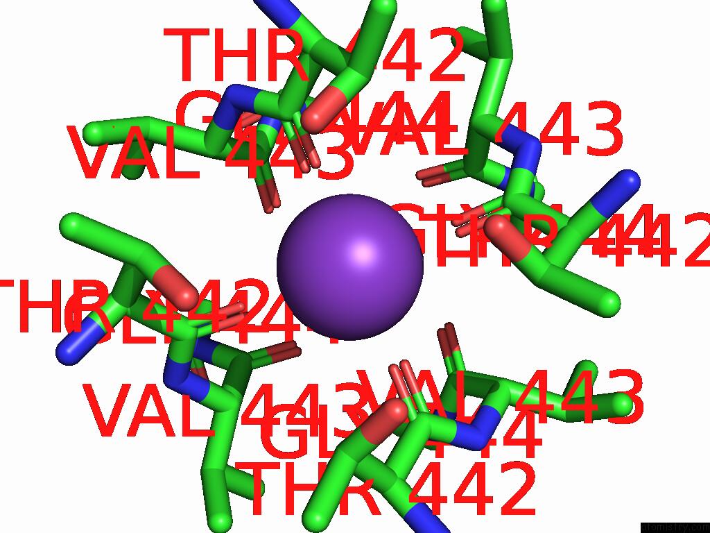
Mono view
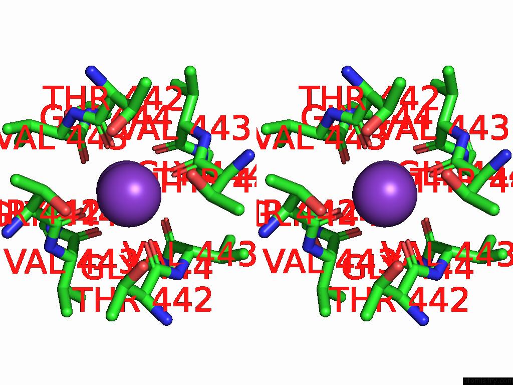
Stereo pair view
Mono view
Stereo pair view
A full contact list of Potassium with other atoms in the K binding
site number 1 of Aca-Ei-Shaker with Free Peptide Conformation A within 5.0Å range:
|
Potassium binding site 2 out of 2 in 9nec
Go back to
Potassium binding site 2 out
of 2 in the Aca-Ei-Shaker with Free Peptide Conformation A
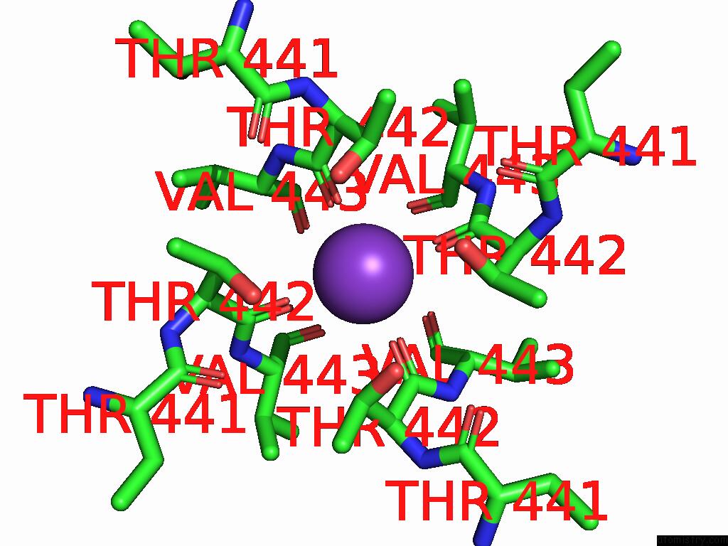
Mono view
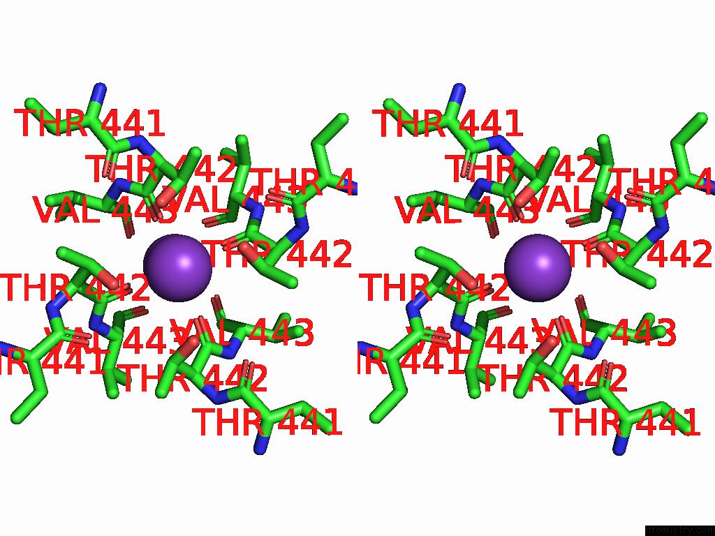
Stereo pair view
Mono view
Stereo pair view
A full contact list of Potassium with other atoms in the K binding
site number 2 of Aca-Ei-Shaker with Free Peptide Conformation A within 5.0Å range:
|
Reference:
X.Tan,
A.I.Fernandez-Marino,
Y.Li,
T.-H.Chang,
K.J.Swartz.
Structural Basis of Fast N-Type Inactivation in Kv Channels Nature 2025.
ISSN: ESSN 1476-4687
DOI: 10.1038/S41586-025-09339-7
Page generated: Sat Aug 23 04:01:43 2025
ISSN: ESSN 1476-4687
DOI: 10.1038/S41586-025-09339-7
Last articles
Mn in 9LJUMn in 9LJW
Mn in 9LJS
Mn in 9LJR
Mn in 9LJT
Mn in 9LJV
Mg in 9UA2
Mg in 9R96
Mg in 9VM1
Mg in 9P01