Potassium »
PDB 7zgt-8b2s »
8axz »
Potassium in PDB 8axz: Crystal Structure of Human Methionine Adenosyltransferase 2A (MAT2A) in Complex with S-Adenosylmethionine, Adenosin and Diphosphono- Aminophosphonic Acid.
Enzymatic activity of Crystal Structure of Human Methionine Adenosyltransferase 2A (MAT2A) in Complex with S-Adenosylmethionine, Adenosin and Diphosphono- Aminophosphonic Acid.
All present enzymatic activity of Crystal Structure of Human Methionine Adenosyltransferase 2A (MAT2A) in Complex with S-Adenosylmethionine, Adenosin and Diphosphono- Aminophosphonic Acid.:
2.5.1.6;
2.5.1.6;
Protein crystallography data
The structure of Crystal Structure of Human Methionine Adenosyltransferase 2A (MAT2A) in Complex with S-Adenosylmethionine, Adenosin and Diphosphono- Aminophosphonic Acid., PDB code: 8axz
was solved by
A.Nawrotek,
L.Vuillard,
L.Miallau,
with X-Ray Crystallography technique. A brief refinement statistics is given in the table below:
| Resolution Low / High (Å) | 58.63 / 1.15 |
| Space group | I 2 2 2 |
| Cell size a, b, c (Å), α, β, γ (°) | 67.554, 94.387, 117.266, 90, 90, 90 |
| R / Rfree (%) | 16.8 / 18.5 |
Other elements in 8axz:
The structure of Crystal Structure of Human Methionine Adenosyltransferase 2A (MAT2A) in Complex with S-Adenosylmethionine, Adenosin and Diphosphono- Aminophosphonic Acid. also contains other interesting chemical elements:
| Magnesium | (Mg) | 2 atoms |
Potassium Binding Sites:
The binding sites of Potassium atom in the Crystal Structure of Human Methionine Adenosyltransferase 2A (MAT2A) in Complex with S-Adenosylmethionine, Adenosin and Diphosphono- Aminophosphonic Acid.
(pdb code 8axz). This binding sites where shown within
5.0 Angstroms radius around Potassium atom.
In total 2 binding sites of Potassium where determined in the Crystal Structure of Human Methionine Adenosyltransferase 2A (MAT2A) in Complex with S-Adenosylmethionine, Adenosin and Diphosphono- Aminophosphonic Acid., PDB code: 8axz:
Jump to Potassium binding site number: 1; 2;
In total 2 binding sites of Potassium where determined in the Crystal Structure of Human Methionine Adenosyltransferase 2A (MAT2A) in Complex with S-Adenosylmethionine, Adenosin and Diphosphono- Aminophosphonic Acid., PDB code: 8axz:
Jump to Potassium binding site number: 1; 2;
Potassium binding site 1 out of 2 in 8axz
Go back to
Potassium binding site 1 out
of 2 in the Crystal Structure of Human Methionine Adenosyltransferase 2A (MAT2A) in Complex with S-Adenosylmethionine, Adenosin and Diphosphono- Aminophosphonic Acid.
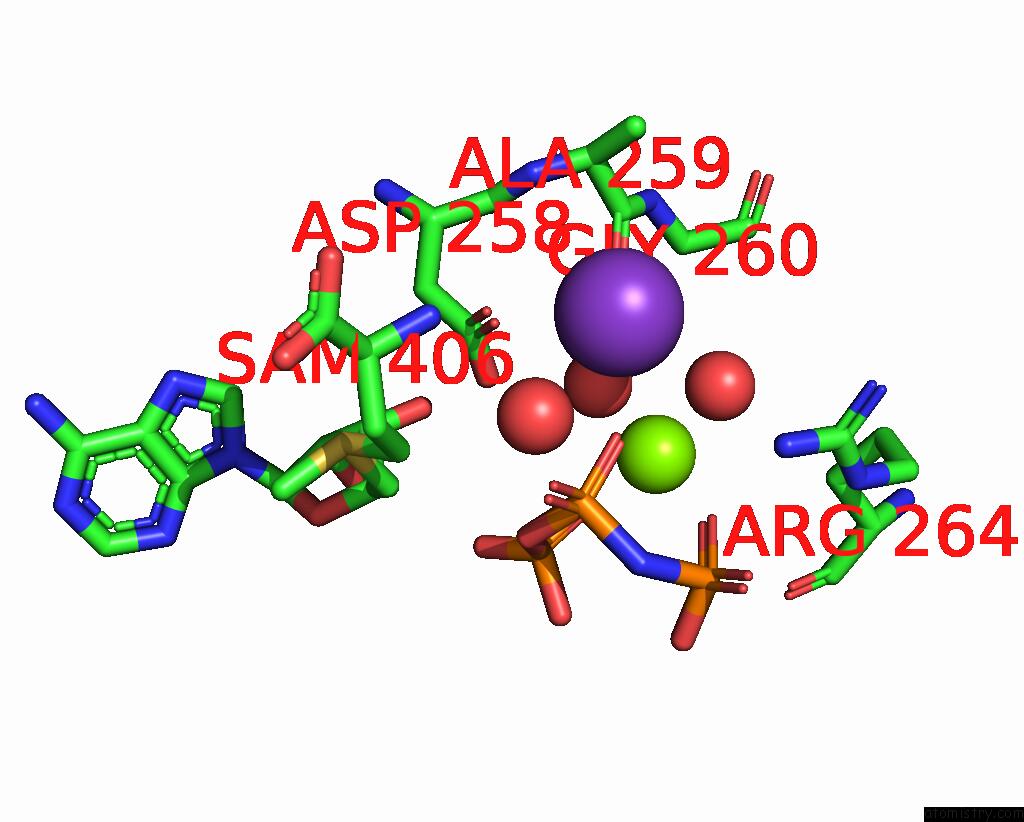
Mono view
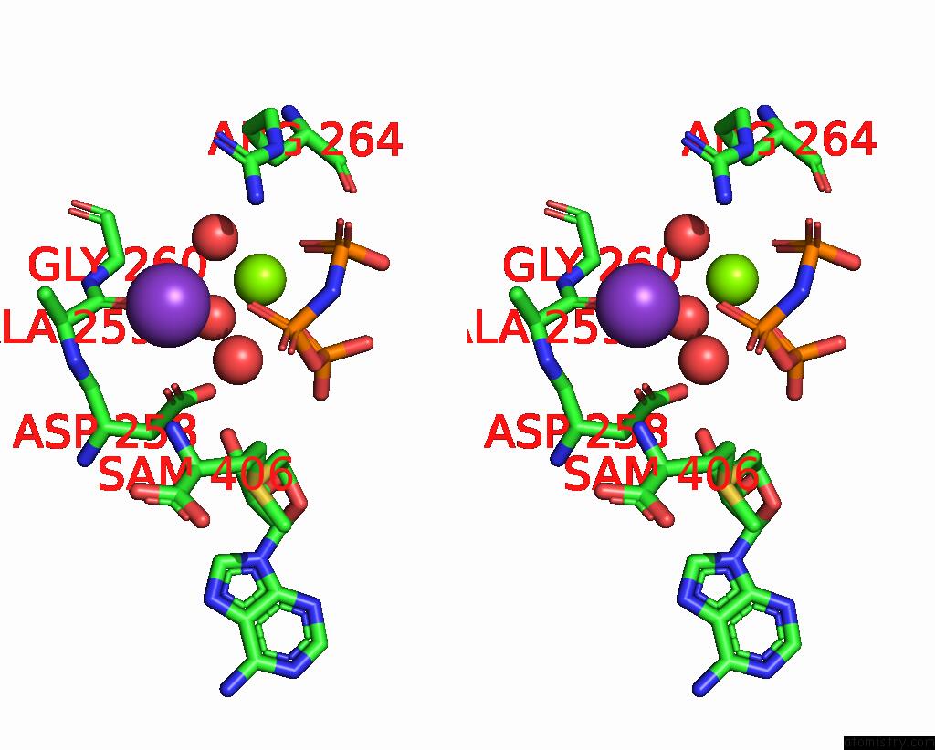
Stereo pair view
Mono view
Stereo pair view
A full contact list of Potassium with other atoms in the K binding
site number 1 of Crystal Structure of Human Methionine Adenosyltransferase 2A (MAT2A) in Complex with S-Adenosylmethionine, Adenosin and Diphosphono- Aminophosphonic Acid. within 5.0Å range:
|
Potassium binding site 2 out of 2 in 8axz
Go back to
Potassium binding site 2 out
of 2 in the Crystal Structure of Human Methionine Adenosyltransferase 2A (MAT2A) in Complex with S-Adenosylmethionine, Adenosin and Diphosphono- Aminophosphonic Acid.
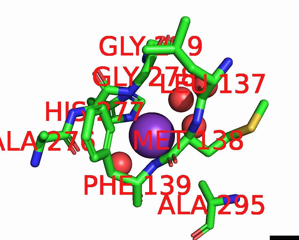
Mono view

Stereo pair view
Mono view
Stereo pair view
A full contact list of Potassium with other atoms in the K binding
site number 2 of Crystal Structure of Human Methionine Adenosyltransferase 2A (MAT2A) in Complex with S-Adenosylmethionine, Adenosin and Diphosphono- Aminophosphonic Acid. within 5.0Å range:
|
Reference:
A.Nawrotek,
L.Vuillard,
L.Miallau.
Crystal Structure of Human Methionine Adenosyltransferase 2A (MAT2A) in Complex with S-Adenosylmethionine, Adenosin and Diphosphono-Aminophosphonic Acid. To Be Published.
Page generated: Sat Aug 9 15:34:25 2025
Last articles
Mg in 1G65Mg in 1G0U
Mg in 1G7T
Mg in 1G75
Mg in 1G6C
Mg in 1G6H
Mg in 1G69
Mg in 1G67
Mg in 1G64
Mg in 1G5T