Potassium »
PDB 7zgt-8b2s »
7zw7 »
Potassium in PDB 7zw7: Crystal Structure of Schistosoma Mansoni HDAC8 in Complex with A Formate Molecule in the Active Site
Protein crystallography data
The structure of Crystal Structure of Schistosoma Mansoni HDAC8 in Complex with A Formate Molecule in the Active Site, PDB code: 7zw7
was solved by
F.Saccoccia,
M.Giannaccari,
G.Ruberti,
with X-Ray Crystallography technique. A brief refinement statistics is given in the table below:
| Resolution Low / High (Å) | 45.89 / 2.85 |
| Space group | P 41 21 2 |
| Cell size a, b, c (Å), α, β, γ (°) | 70.957, 70.957, 180.514, 90, 90, 90 |
| R / Rfree (%) | 23.4 / 27.5 |
Other elements in 7zw7:
The structure of Crystal Structure of Schistosoma Mansoni HDAC8 in Complex with A Formate Molecule in the Active Site also contains other interesting chemical elements:
| Zinc | (Zn) | 1 atom |
| Chlorine | (Cl) | 1 atom |
Potassium Binding Sites:
The binding sites of Potassium atom in the Crystal Structure of Schistosoma Mansoni HDAC8 in Complex with A Formate Molecule in the Active Site
(pdb code 7zw7). This binding sites where shown within
5.0 Angstroms radius around Potassium atom.
In total 2 binding sites of Potassium where determined in the Crystal Structure of Schistosoma Mansoni HDAC8 in Complex with A Formate Molecule in the Active Site, PDB code: 7zw7:
Jump to Potassium binding site number: 1; 2;
In total 2 binding sites of Potassium where determined in the Crystal Structure of Schistosoma Mansoni HDAC8 in Complex with A Formate Molecule in the Active Site, PDB code: 7zw7:
Jump to Potassium binding site number: 1; 2;
Potassium binding site 1 out of 2 in 7zw7
Go back to
Potassium binding site 1 out
of 2 in the Crystal Structure of Schistosoma Mansoni HDAC8 in Complex with A Formate Molecule in the Active Site
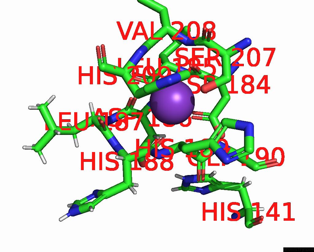
Mono view
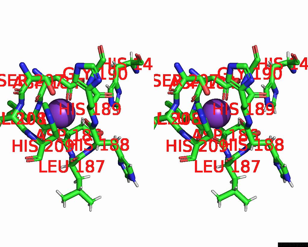
Stereo pair view
Mono view
Stereo pair view
A full contact list of Potassium with other atoms in the K binding
site number 1 of Crystal Structure of Schistosoma Mansoni HDAC8 in Complex with A Formate Molecule in the Active Site within 5.0Å range:
|
Potassium binding site 2 out of 2 in 7zw7
Go back to
Potassium binding site 2 out
of 2 in the Crystal Structure of Schistosoma Mansoni HDAC8 in Complex with A Formate Molecule in the Active Site
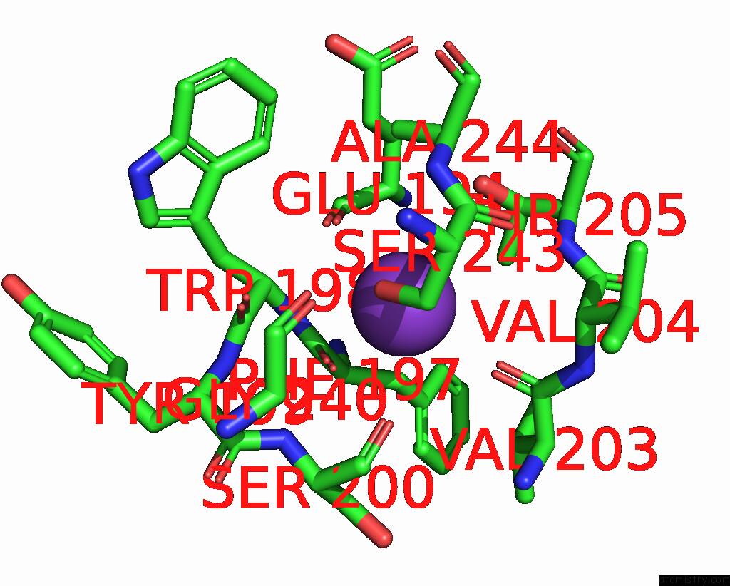
Mono view
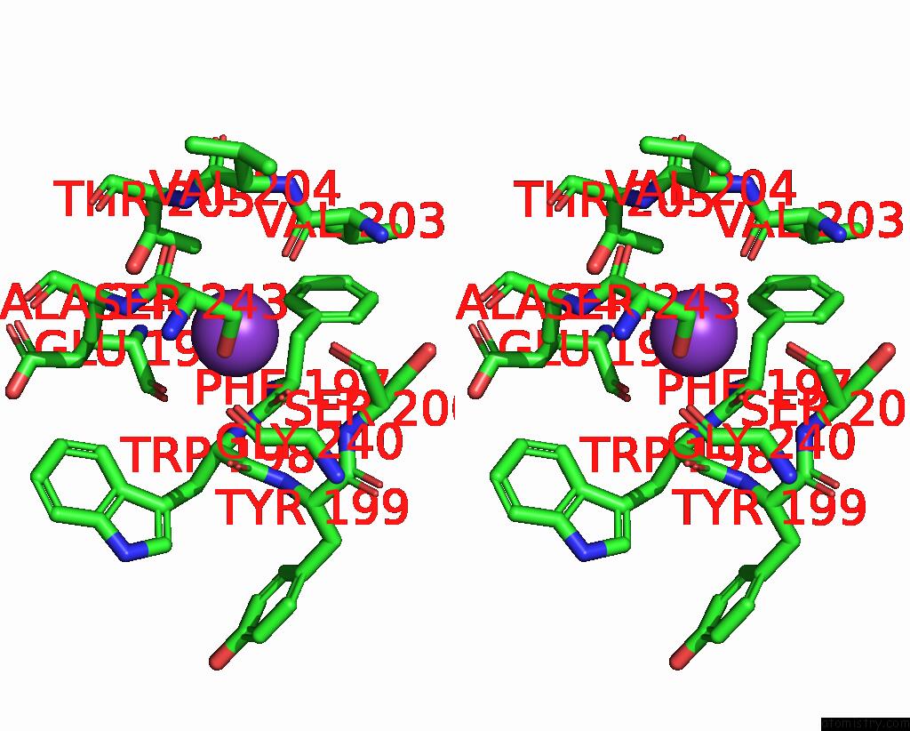
Stereo pair view
Mono view
Stereo pair view
A full contact list of Potassium with other atoms in the K binding
site number 2 of Crystal Structure of Schistosoma Mansoni HDAC8 in Complex with A Formate Molecule in the Active Site within 5.0Å range:
|
Reference:
F.Saccoccia,
M.Giannaccari,
G.Ruberti.
Crystal Structure of Schistosoma Mansoni HDAC8 in Complex with A Formate Molecule in the Active Site To Be Published.
Page generated: Sat Aug 9 15:32:17 2025
Last articles
Mn in 3FVMMn in 3FQ5
Mn in 3FM1
Mn in 3FJQ
Mn in 3FJW
Mn in 3FGO
Mn in 3FHI
Mn in 3FGZ
Mn in 3FGA
Mn in 3FFX