Potassium »
PDB 7xxk-7zgo »
7zd4 »
Potassium in PDB 7zd4: Crystal Structure of Pseudomonas Aeruginosa S-Adenosyl-L-Homocysteine Hydrolase Soaked with Cu+ Ions
Enzymatic activity of Crystal Structure of Pseudomonas Aeruginosa S-Adenosyl-L-Homocysteine Hydrolase Soaked with Cu+ Ions
All present enzymatic activity of Crystal Structure of Pseudomonas Aeruginosa S-Adenosyl-L-Homocysteine Hydrolase Soaked with Cu+ Ions:
3.3.1.1;
3.3.1.1;
Protein crystallography data
The structure of Crystal Structure of Pseudomonas Aeruginosa S-Adenosyl-L-Homocysteine Hydrolase Soaked with Cu+ Ions, PDB code: 7zd4
was solved by
P.H.Malecki,
M.Gawel,
K.Brzezinski,
with X-Ray Crystallography technique. A brief refinement statistics is given in the table below:
| Resolution Low / High (Å) | 35.29 / 2.14 |
| Space group | C 1 2 1 |
| Cell size a, b, c (Å), α, β, γ (°) | 175.68, 133.57, 107.04, 90, 105.22, 90 |
| R / Rfree (%) | 18.3 / 22.7 |
Other elements in 7zd4:
The structure of Crystal Structure of Pseudomonas Aeruginosa S-Adenosyl-L-Homocysteine Hydrolase Soaked with Cu+ Ions also contains other interesting chemical elements:
| Bromine | (Br) | 1 atom |
Potassium Binding Sites:
The binding sites of Potassium atom in the Crystal Structure of Pseudomonas Aeruginosa S-Adenosyl-L-Homocysteine Hydrolase Soaked with Cu+ Ions
(pdb code 7zd4). This binding sites where shown within
5.0 Angstroms radius around Potassium atom.
In total 4 binding sites of Potassium where determined in the Crystal Structure of Pseudomonas Aeruginosa S-Adenosyl-L-Homocysteine Hydrolase Soaked with Cu+ Ions, PDB code: 7zd4:
Jump to Potassium binding site number: 1; 2; 3; 4;
In total 4 binding sites of Potassium where determined in the Crystal Structure of Pseudomonas Aeruginosa S-Adenosyl-L-Homocysteine Hydrolase Soaked with Cu+ Ions, PDB code: 7zd4:
Jump to Potassium binding site number: 1; 2; 3; 4;
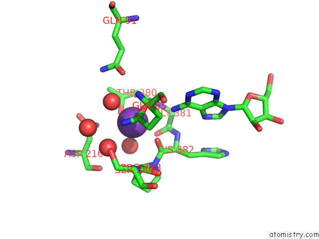
Potassium binding site 1 out of 4 in 7zd4
Go back to
Potassium binding site 1 out
of 4 in the Crystal Structure of Pseudomonas Aeruginosa S-Adenosyl-L-Homocysteine Hydrolase Soaked with Cu+ Ions
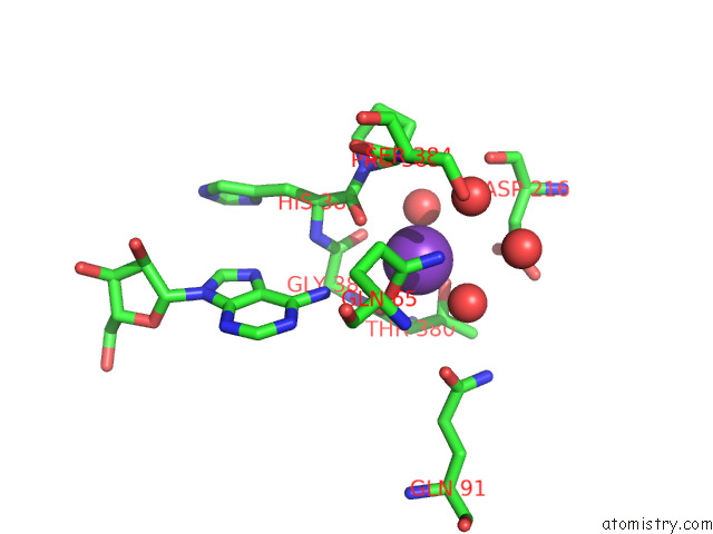
Mono view
Stereo pair view
Mono view
Stereo pair view
A full contact list of Potassium with other atoms in the K binding
site number 1 of Crystal Structure of Pseudomonas Aeruginosa S-Adenosyl-L-Homocysteine Hydrolase Soaked with Cu+ Ions within 5.0Å range:
|
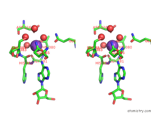
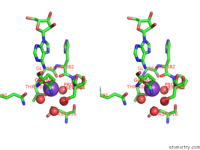
Potassium binding site 2 out of 4 in 7zd4
Go back to
Potassium binding site 2 out
of 4 in the Crystal Structure of Pseudomonas Aeruginosa S-Adenosyl-L-Homocysteine Hydrolase Soaked with Cu+ Ions
Mono view
Stereo pair view
Mono view
Stereo pair view
A full contact list of Potassium with other atoms in the K binding
site number 2 of Crystal Structure of Pseudomonas Aeruginosa S-Adenosyl-L-Homocysteine Hydrolase Soaked with Cu+ Ions within 5.0Å range:
|
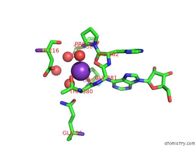
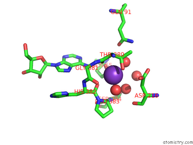
Potassium binding site 3 out of 4 in 7zd4
Go back to
Potassium binding site 3 out
of 4 in the Crystal Structure of Pseudomonas Aeruginosa S-Adenosyl-L-Homocysteine Hydrolase Soaked with Cu+ Ions
Mono view
Stereo pair view
Mono view
Stereo pair view
A full contact list of Potassium with other atoms in the K binding
site number 3 of Crystal Structure of Pseudomonas Aeruginosa S-Adenosyl-L-Homocysteine Hydrolase Soaked with Cu+ Ions within 5.0Å range:
|
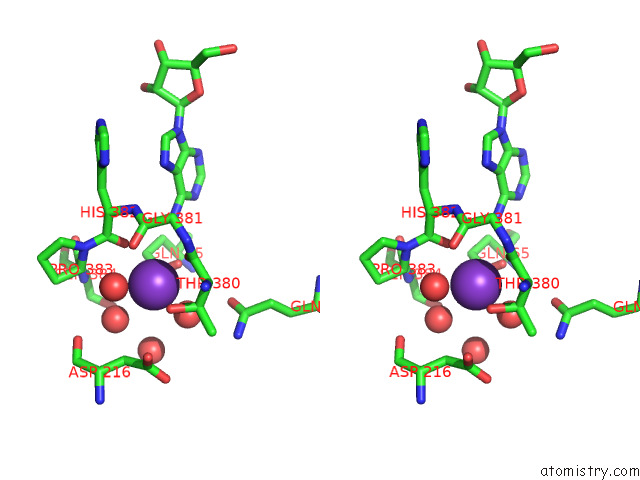
Potassium binding site 4 out of 4 in 7zd4
Go back to
Potassium binding site 4 out
of 4 in the Crystal Structure of Pseudomonas Aeruginosa S-Adenosyl-L-Homocysteine Hydrolase Soaked with Cu+ Ions
Mono view
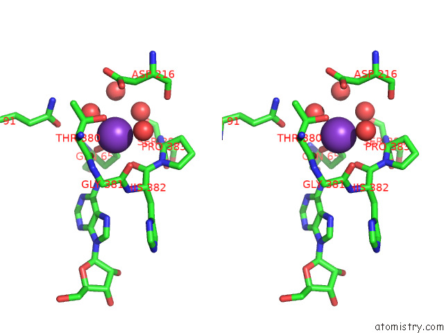
Stereo pair view
Mono view
Stereo pair view
A full contact list of Potassium with other atoms in the K binding
site number 4 of Crystal Structure of Pseudomonas Aeruginosa S-Adenosyl-L-Homocysteine Hydrolase Soaked with Cu+ Ions within 5.0Å range:
|
Reference:
P.H.Malecki,
M.Gawel,
K.Brzezinski.
Crystal Structure of Pseudomonas Aeruginosa S-Adenosyl-L-Homocysteine Hydrolase Soaked with Cu+ Ions To Be Published.
Page generated: Sat Aug 9 15:23:50 2025
Last articles
Mg in 6LU1Mg in 6LT4
Mg in 6LY7
Mg in 6LY6
Mg in 6LY3
Mg in 6LX1
Mg in 6LTW
Mg in 6LVW
Mg in 6LUH
Mg in 6LTS