Potassium »
PDB 7uut-7xx6 »
7wik »
Potassium in PDB 7wik: Crystal Structure of Oligoribonuclease of Mycobacterium Smegmatis MC2 155
Enzymatic activity of Crystal Structure of Oligoribonuclease of Mycobacterium Smegmatis MC2 155
All present enzymatic activity of Crystal Structure of Oligoribonuclease of Mycobacterium Smegmatis MC2 155:
3.1.13.3;
3.1.13.3;
Protein crystallography data
The structure of Crystal Structure of Oligoribonuclease of Mycobacterium Smegmatis MC2 155, PDB code: 7wik
was solved by
P.Badhwar,
B.Taneja,
with X-Ray Crystallography technique. A brief refinement statistics is given in the table below:
| Resolution Low / High (Å) | 81.07 / 1.87 |
| Space group | P 21 21 21 |
| Cell size a, b, c (Å), α, β, γ (°) | 60.25, 97.26, 146.73, 90, 90, 90 |
| R / Rfree (%) | 17.9 / 22.2 |
Other elements in 7wik:
The structure of Crystal Structure of Oligoribonuclease of Mycobacterium Smegmatis MC2 155 also contains other interesting chemical elements:
| Magnesium | (Mg) | 1 atom |
Potassium Binding Sites:
The binding sites of Potassium atom in the Crystal Structure of Oligoribonuclease of Mycobacterium Smegmatis MC2 155
(pdb code 7wik). This binding sites where shown within
5.0 Angstroms radius around Potassium atom.
In total 4 binding sites of Potassium where determined in the Crystal Structure of Oligoribonuclease of Mycobacterium Smegmatis MC2 155, PDB code: 7wik:
Jump to Potassium binding site number: 1; 2; 3; 4;
In total 4 binding sites of Potassium where determined in the Crystal Structure of Oligoribonuclease of Mycobacterium Smegmatis MC2 155, PDB code: 7wik:
Jump to Potassium binding site number: 1; 2; 3; 4;
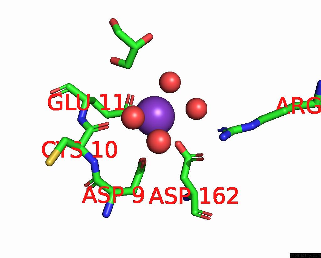
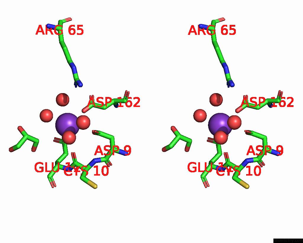
Potassium binding site 1 out of 4 in 7wik
Go back to
Potassium binding site 1 out
of 4 in the Crystal Structure of Oligoribonuclease of Mycobacterium Smegmatis MC2 155
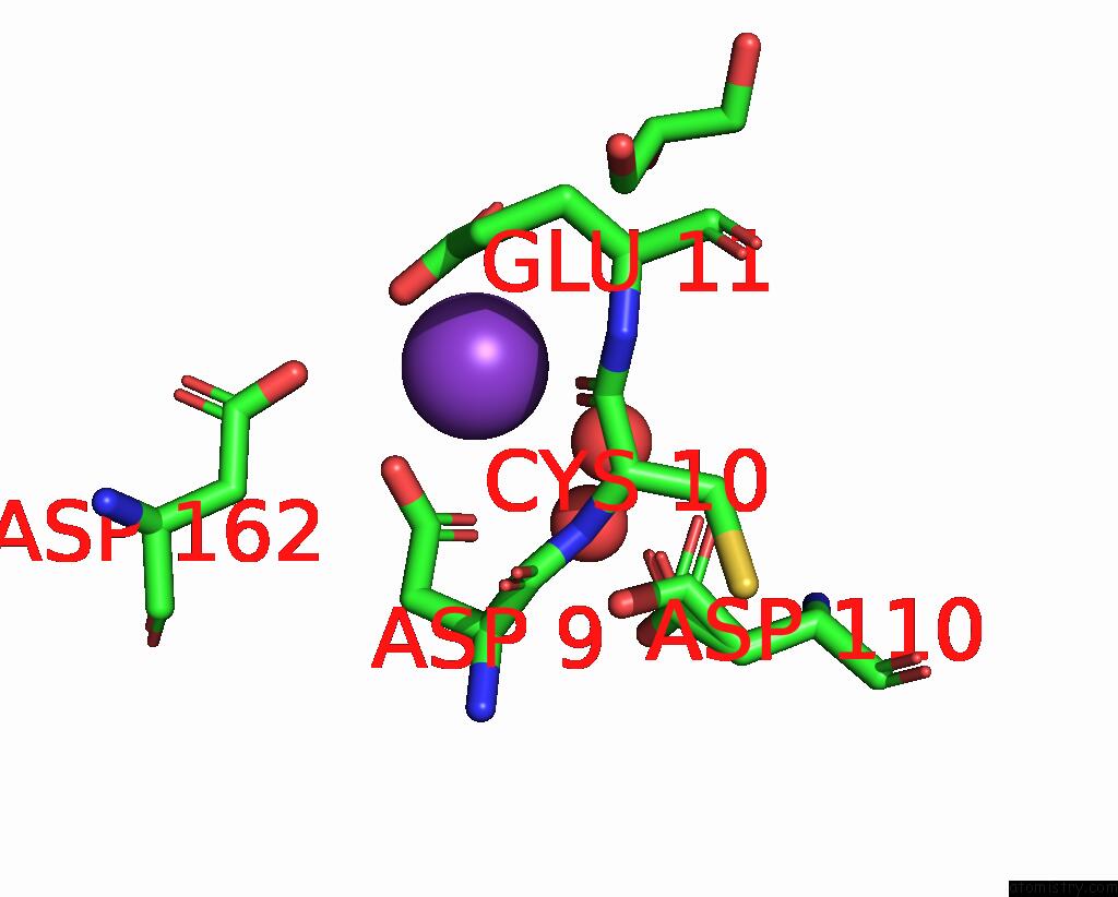
Mono view
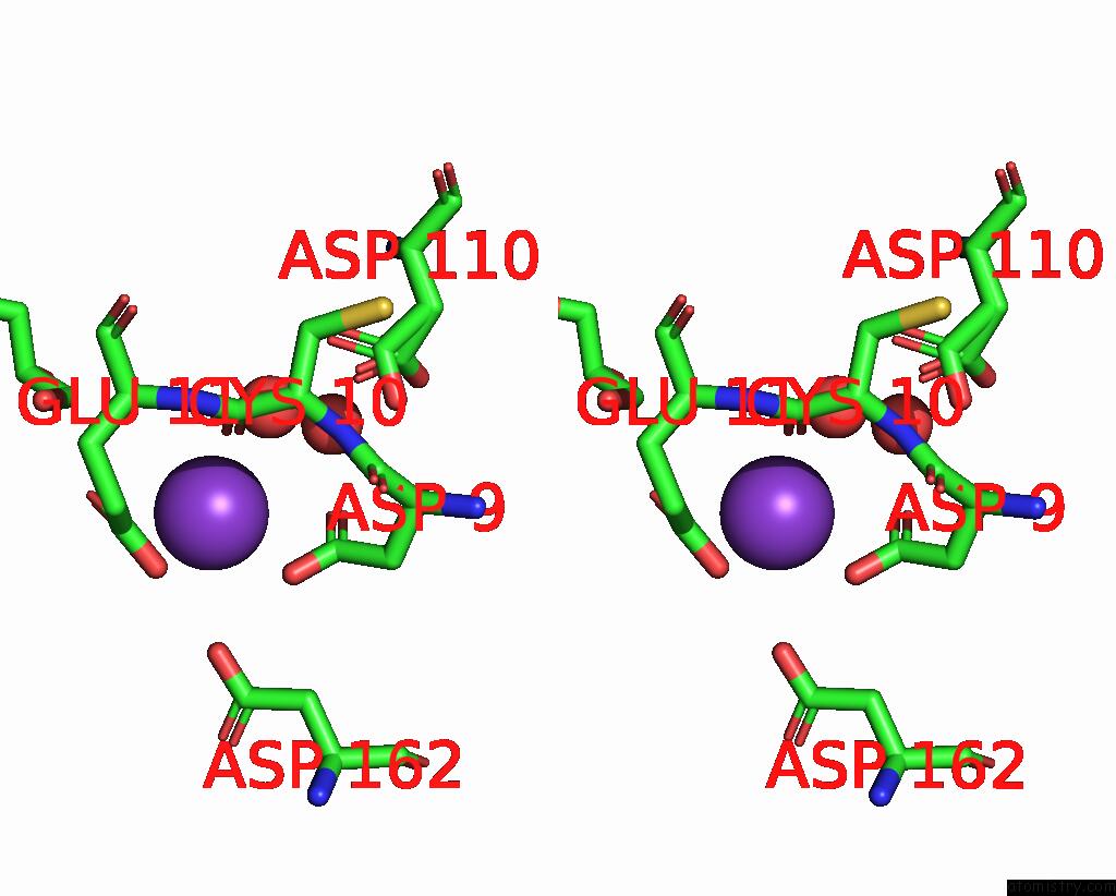
Stereo pair view
Mono view
Stereo pair view
A full contact list of Potassium with other atoms in the K binding
site number 1 of Crystal Structure of Oligoribonuclease of Mycobacterium Smegmatis MC2 155 within 5.0Å range:
|
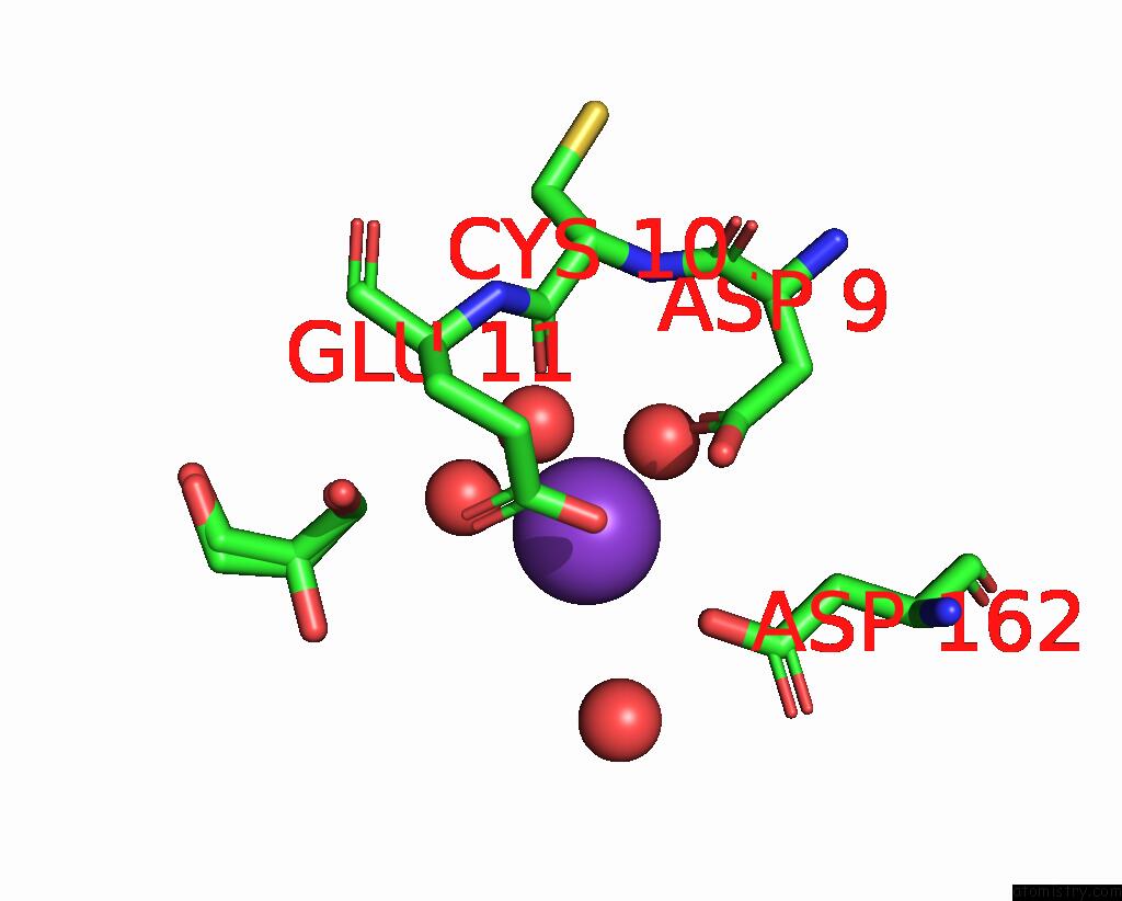
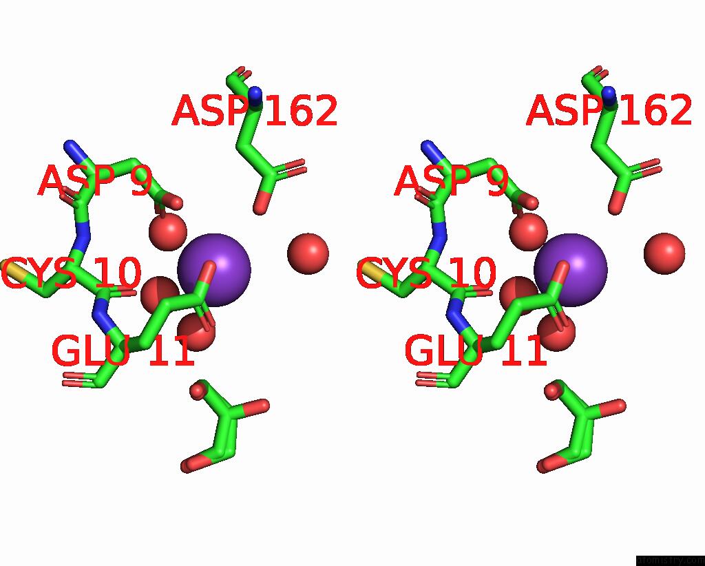
Potassium binding site 2 out of 4 in 7wik
Go back to
Potassium binding site 2 out
of 4 in the Crystal Structure of Oligoribonuclease of Mycobacterium Smegmatis MC2 155
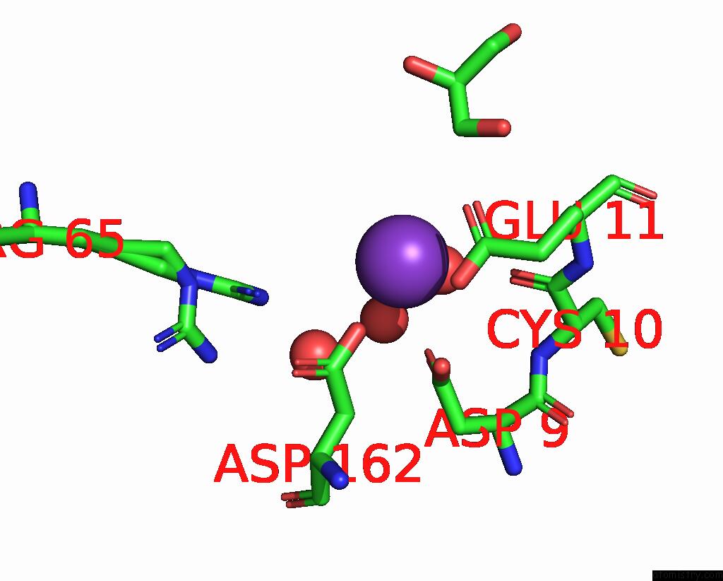
Mono view
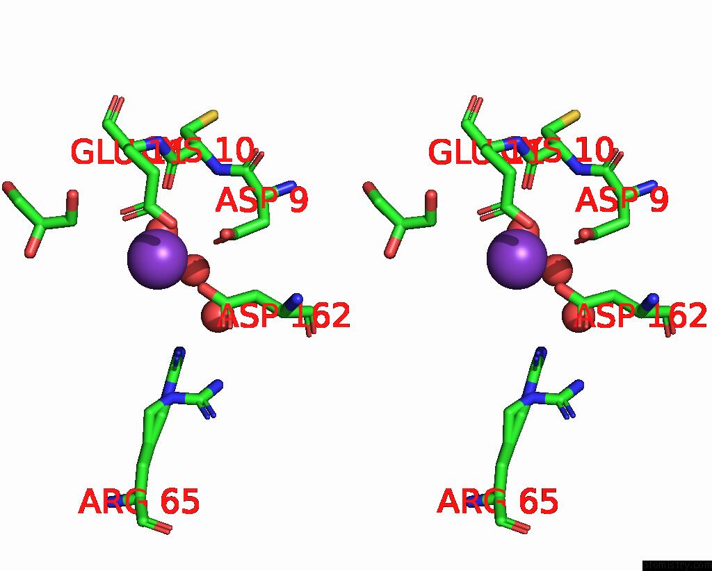
Stereo pair view
Mono view
Stereo pair view
A full contact list of Potassium with other atoms in the K binding
site number 2 of Crystal Structure of Oligoribonuclease of Mycobacterium Smegmatis MC2 155 within 5.0Å range:
|
Potassium binding site 3 out of 4 in 7wik
Go back to
Potassium binding site 3 out
of 4 in the Crystal Structure of Oligoribonuclease of Mycobacterium Smegmatis MC2 155
Mono view
Stereo pair view
Mono view
Stereo pair view
A full contact list of Potassium with other atoms in the K binding
site number 3 of Crystal Structure of Oligoribonuclease of Mycobacterium Smegmatis MC2 155 within 5.0Å range:
|
Potassium binding site 4 out of 4 in 7wik
Go back to
Potassium binding site 4 out
of 4 in the Crystal Structure of Oligoribonuclease of Mycobacterium Smegmatis MC2 155
Mono view
Stereo pair view
Mono view
Stereo pair view
A full contact list of Potassium with other atoms in the K binding
site number 4 of Crystal Structure of Oligoribonuclease of Mycobacterium Smegmatis MC2 155 within 5.0Å range:
|
Reference:
P.Badhwar,
S.H.Khan,
B.Taneja.
Three-Dimensional Structure of A Mycobacterial Oligoribonuclease Reveals A Unique C-Terminal Tail That Stabilizes the Homodimer. J.Biol.Chem. V. 298 02595 2022.
ISSN: ESSN 1083-351X
PubMed: 36244449
DOI: 10.1016/J.JBC.2022.102595
Page generated: Sat Aug 9 15:10:05 2025
ISSN: ESSN 1083-351X
PubMed: 36244449
DOI: 10.1016/J.JBC.2022.102595
Last articles
Mg in 5M70Mg in 5M6Z
Mg in 5M5O
Mg in 5M5N
Mg in 5M5L
Mg in 5M5M
Mg in 5M45
Mg in 5M5I
Mg in 5M5C
Mg in 5M4L