Potassium »
PDB 7uut-7xx6 »
7w93 »
Potassium in PDB 7w93: Crystal Structure of E.Coli Pseudouridine Kinase Psuk Complexed with N1-Methyl-Pseudouridine
Protein crystallography data
The structure of Crystal Structure of E.Coli Pseudouridine Kinase Psuk Complexed with N1-Methyl-Pseudouridine, PDB code: 7w93
was solved by
K.J.Li,
X.J.Li,
B.X.Wu,
with X-Ray Crystallography technique. A brief refinement statistics is given in the table below:
| Resolution Low / High (Å) | 29.19 / 1.90 |
| Space group | P 63 2 2 |
| Cell size a, b, c (Å), α, β, γ (°) | 185.83, 185.83, 52.51, 90, 90, 120 |
| R / Rfree (%) | 19.6 / 21.5 |
Potassium Binding Sites:
The binding sites of Potassium atom in the Crystal Structure of E.Coli Pseudouridine Kinase Psuk Complexed with N1-Methyl-Pseudouridine
(pdb code 7w93). This binding sites where shown within
5.0 Angstroms radius around Potassium atom.
In total only one binding site of Potassium was determined in the Crystal Structure of E.Coli Pseudouridine Kinase Psuk Complexed with N1-Methyl-Pseudouridine, PDB code: 7w93:
In total only one binding site of Potassium was determined in the Crystal Structure of E.Coli Pseudouridine Kinase Psuk Complexed with N1-Methyl-Pseudouridine, PDB code: 7w93:
Potassium binding site 1 out of 1 in 7w93
Go back to
Potassium binding site 1 out
of 1 in the Crystal Structure of E.Coli Pseudouridine Kinase Psuk Complexed with N1-Methyl-Pseudouridine
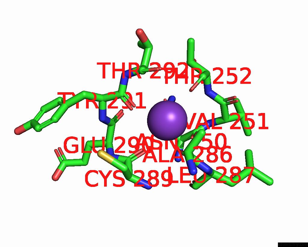
Mono view
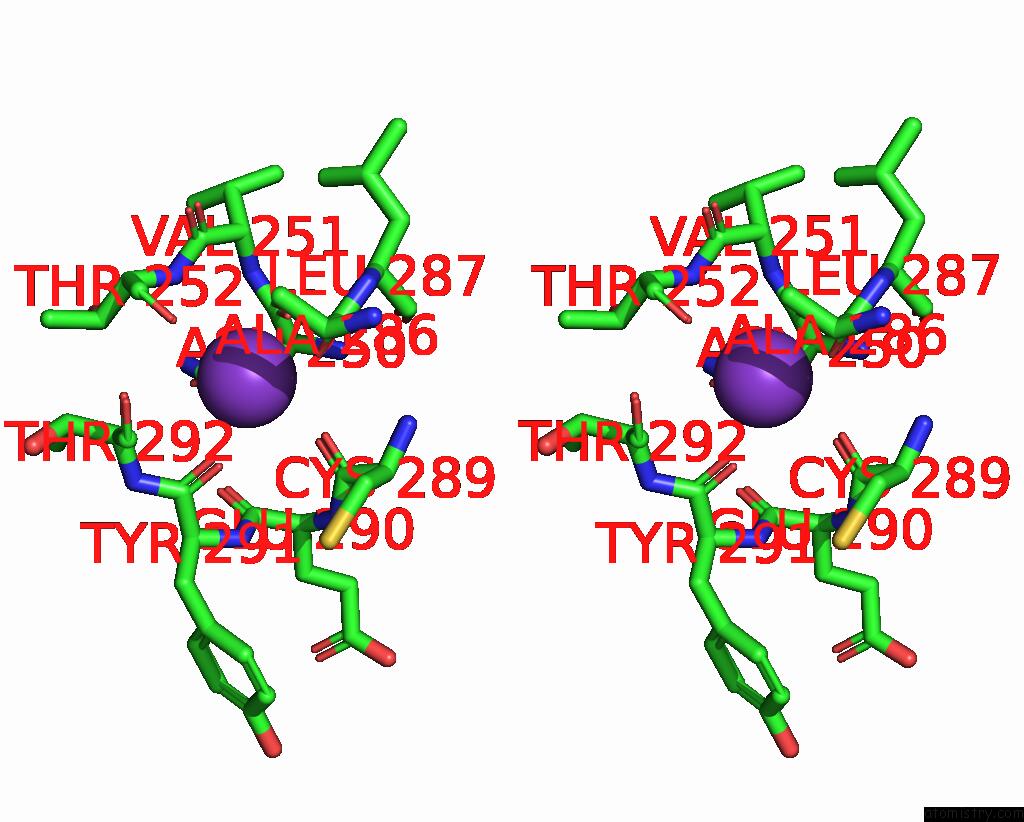
Stereo pair view
Mono view
Stereo pair view
A full contact list of Potassium with other atoms in the K binding
site number 1 of Crystal Structure of E.Coli Pseudouridine Kinase Psuk Complexed with N1-Methyl-Pseudouridine within 5.0Å range:
|
Reference:
X.Li,
K.Li,
W.Guo,
Y.Wen,
C.Meng,
B.Wu.
Structure Characterization of Escherichia Coli Pseudouridine Kinase Psuk. Front Microbiol V. 13 26099 2022.
ISSN: ESSN 1664-302X
PubMed: 35783380
DOI: 10.3389/FMICB.2022.926099
Page generated: Sat Aug 9 15:09:42 2025
ISSN: ESSN 1664-302X
PubMed: 35783380
DOI: 10.3389/FMICB.2022.926099
Last articles
Mg in 4DPGMg in 4DQP
Mg in 4DQQ
Mg in 4DPM
Mg in 4DPV
Mg in 4DQI
Mg in 4DOB
Mg in 4DOC
Mg in 4DMZ
Mg in 4DOA