Potassium »
PDB 7ruu-7ta6 »
7sev »
Potassium in PDB 7sev: Crystal Structure of E Coli Contaminant Protein Yadf Co-Purified with A Plant Protein
Enzymatic activity of Crystal Structure of E Coli Contaminant Protein Yadf Co-Purified with A Plant Protein
All present enzymatic activity of Crystal Structure of E Coli Contaminant Protein Yadf Co-Purified with A Plant Protein:
4.2.1.1;
4.2.1.1;
Protein crystallography data
The structure of Crystal Structure of E Coli Contaminant Protein Yadf Co-Purified with A Plant Protein, PDB code: 7sev
was solved by
L.Chai,
P.Zhu,
J.Chai,
C.Pang,
B.Andi,
S.Mcsweeney,
J.Shanklin,
Q.Liu,
with X-Ray Crystallography technique. A brief refinement statistics is given in the table below:
| Resolution Low / High (Å) | 36.03 / 2.30 |
| Space group | P 42 21 2 |
| Cell size a, b, c (Å), α, β, γ (°) | 67.5, 67.5, 85.212, 90, 90, 90 |
| R / Rfree (%) | 20.4 / 24.3 |
Other elements in 7sev:
The structure of Crystal Structure of E Coli Contaminant Protein Yadf Co-Purified with A Plant Protein also contains other interesting chemical elements:
| Zinc | (Zn) | 1 atom |
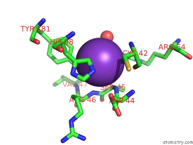
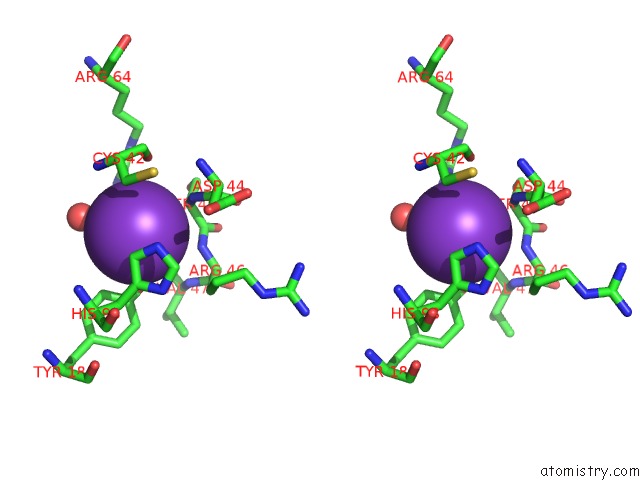
Potassium Binding Sites:
The binding sites of Potassium atom in the Crystal Structure of E Coli Contaminant Protein Yadf Co-Purified with A Plant Protein
(pdb code 7sev). This binding sites where shown within
5.0 Angstroms radius around Potassium atom.
In total only one binding site of Potassium was determined in the Crystal Structure of E Coli Contaminant Protein Yadf Co-Purified with A Plant Protein, PDB code: 7sev:
In total only one binding site of Potassium was determined in the Crystal Structure of E Coli Contaminant Protein Yadf Co-Purified with A Plant Protein, PDB code: 7sev:
Potassium binding site 1 out of 1 in 7sev
Go back to
Potassium binding site 1 out
of 1 in the Crystal Structure of E Coli Contaminant Protein Yadf Co-Purified with A Plant Protein
Mono view
Stereo pair view
Mono view
Stereo pair view
A full contact list of Potassium with other atoms in the K binding
site number 1 of Crystal Structure of E Coli Contaminant Protein Yadf Co-Purified with A Plant Protein within 5.0Å range:
|
Reference:
L.Chai,
P.Zhu,
J.Chai,
C.Pang,
B.Andi,
S.Mcsweeney,
J.Shanklin,
Q.Liu.
Alphafold Protein Structure Database For Sequence-Independent Molecular Replacement To Be Published.
Page generated: Sat Aug 9 14:52:11 2025
Last articles
Mg in 9L14Mg in 9KUK
Mg in 9KUM
Mg in 9KUL
Mg in 9KQP
Mg in 9KQB
Mg in 9K3Q
Mg in 9KOK
Mg in 9KBJ
Mg in 9KLN