Potassium »
PDB 7qr3-7rut »
7qzj »
Potassium in PDB 7qzj: 1.55 A X-Ray Crystallographic Structure of Saph From Streptomyces Sp. (HPH0547) Involved in Pseudouridimycin Biosynthesis
Protein crystallography data
The structure of 1.55 A X-Ray Crystallographic Structure of Saph From Streptomyces Sp. (HPH0547) Involved in Pseudouridimycin Biosynthesis, PDB code: 7qzj
was solved by
R.Schnell,
G.Schneider,
with X-Ray Crystallography technique. A brief refinement statistics is given in the table below:
| Resolution Low / High (Å) | 44.47 / 1.55 |
| Space group | P 21 21 21 |
| Cell size a, b, c (Å), α, β, γ (°) | 62.581, 63.191, 211.415, 90, 90, 90 |
| R / Rfree (%) | 16 / 18.2 |
Potassium Binding Sites:
The binding sites of Potassium atom in the 1.55 A X-Ray Crystallographic Structure of Saph From Streptomyces Sp. (HPH0547) Involved in Pseudouridimycin Biosynthesis
(pdb code 7qzj). This binding sites where shown within
5.0 Angstroms radius around Potassium atom.
In total 3 binding sites of Potassium where determined in the 1.55 A X-Ray Crystallographic Structure of Saph From Streptomyces Sp. (HPH0547) Involved in Pseudouridimycin Biosynthesis, PDB code: 7qzj:
Jump to Potassium binding site number: 1; 2; 3;
In total 3 binding sites of Potassium where determined in the 1.55 A X-Ray Crystallographic Structure of Saph From Streptomyces Sp. (HPH0547) Involved in Pseudouridimycin Biosynthesis, PDB code: 7qzj:
Jump to Potassium binding site number: 1; 2; 3;
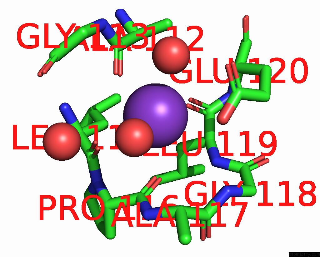
Potassium binding site 1 out of 3 in 7qzj
Go back to
Potassium binding site 1 out
of 3 in the 1.55 A X-Ray Crystallographic Structure of Saph From Streptomyces Sp. (HPH0547) Involved in Pseudouridimycin Biosynthesis

Mono view
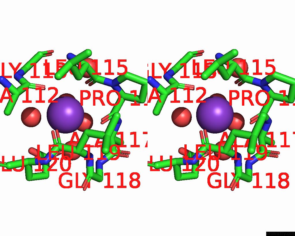
Stereo pair view
Mono view
Stereo pair view
A full contact list of Potassium with other atoms in the K binding
site number 1 of 1.55 A X-Ray Crystallographic Structure of Saph From Streptomyces Sp. (HPH0547) Involved in Pseudouridimycin Biosynthesis within 5.0Å range:
|
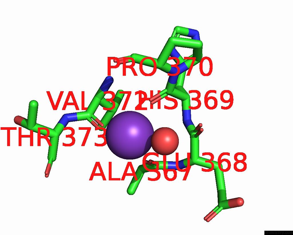
Potassium binding site 2 out of 3 in 7qzj
Go back to
Potassium binding site 2 out
of 3 in the 1.55 A X-Ray Crystallographic Structure of Saph From Streptomyces Sp. (HPH0547) Involved in Pseudouridimycin Biosynthesis
Mono view

Stereo pair view
Mono view
Stereo pair view
A full contact list of Potassium with other atoms in the K binding
site number 2 of 1.55 A X-Ray Crystallographic Structure of Saph From Streptomyces Sp. (HPH0547) Involved in Pseudouridimycin Biosynthesis within 5.0Å range:
|
Potassium binding site 3 out of 3 in 7qzj
Go back to
Potassium binding site 3 out
of 3 in the 1.55 A X-Ray Crystallographic Structure of Saph From Streptomyces Sp. (HPH0547) Involved in Pseudouridimycin Biosynthesis
Mono view

Stereo pair view
Mono view
Stereo pair view
A full contact list of Potassium with other atoms in the K binding
site number 3 of 1.55 A X-Ray Crystallographic Structure of Saph From Streptomyces Sp. (HPH0547) Involved in Pseudouridimycin Biosynthesis within 5.0Å range:
|
Reference:
R.Schnell,
G.Schneider.
1.55 A X-Ray Crystallographic Structure of Saph From Streptomyces Sp. (HPH0547) Involved in Pseudouridimycin Biosynthesis To Be Published.
Page generated: Sat Aug 9 14:40:08 2025
Last articles
Mg in 6KF3Mg in 6KE4
Mg in 6KE0
Mg in 6KDZ
Mg in 6KDX
Mg in 6KDN
Mg in 6KDM
Mg in 6KDK
Mg in 6KDJ
Mg in 6KD2