Potassium »
PDB 7jvv-7kw7 »
7kut »
Potassium in PDB 7kut: Crystal Structure of Danio Rerio Histone Deacetylase 10 H137A Mutant in Complex with N-Acetylputrescine (Tetrahedral Intermediate)
Enzymatic activity of Crystal Structure of Danio Rerio Histone Deacetylase 10 H137A Mutant in Complex with N-Acetylputrescine (Tetrahedral Intermediate)
All present enzymatic activity of Crystal Structure of Danio Rerio Histone Deacetylase 10 H137A Mutant in Complex with N-Acetylputrescine (Tetrahedral Intermediate):
3.5.1.48; 3.5.1.62;
3.5.1.48; 3.5.1.62;
Protein crystallography data
The structure of Crystal Structure of Danio Rerio Histone Deacetylase 10 H137A Mutant in Complex with N-Acetylputrescine (Tetrahedral Intermediate), PDB code: 7kut
was solved by
C.J.Herbst-Gervasoni,
D.W.Christianson,
with X-Ray Crystallography technique. A brief refinement statistics is given in the table below:
| Resolution Low / High (Å) | 60.50 / 2.05 |
| Space group | P 31 2 1 |
| Cell size a, b, c (Å), α, β, γ (°) | 80.248, 80.248, 245.96, 90, 90, 120 |
| R / Rfree (%) | 17.6 / 21.9 |
Other elements in 7kut:
The structure of Crystal Structure of Danio Rerio Histone Deacetylase 10 H137A Mutant in Complex with N-Acetylputrescine (Tetrahedral Intermediate) also contains other interesting chemical elements:
| Sodium | (Na) | 1 atom |
| Zinc | (Zn) | 1 atom |
Potassium Binding Sites:
The binding sites of Potassium atom in the Crystal Structure of Danio Rerio Histone Deacetylase 10 H137A Mutant in Complex with N-Acetylputrescine (Tetrahedral Intermediate)
(pdb code 7kut). This binding sites where shown within
5.0 Angstroms radius around Potassium atom.
In total 2 binding sites of Potassium where determined in the Crystal Structure of Danio Rerio Histone Deacetylase 10 H137A Mutant in Complex with N-Acetylputrescine (Tetrahedral Intermediate), PDB code: 7kut:
Jump to Potassium binding site number: 1; 2;
In total 2 binding sites of Potassium where determined in the Crystal Structure of Danio Rerio Histone Deacetylase 10 H137A Mutant in Complex with N-Acetylputrescine (Tetrahedral Intermediate), PDB code: 7kut:
Jump to Potassium binding site number: 1; 2;
Potassium binding site 1 out of 2 in 7kut
Go back to
Potassium binding site 1 out
of 2 in the Crystal Structure of Danio Rerio Histone Deacetylase 10 H137A Mutant in Complex with N-Acetylputrescine (Tetrahedral Intermediate)
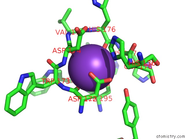
Mono view
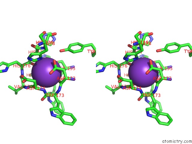
Stereo pair view
Mono view
Stereo pair view
A full contact list of Potassium with other atoms in the K binding
site number 1 of Crystal Structure of Danio Rerio Histone Deacetylase 10 H137A Mutant in Complex with N-Acetylputrescine (Tetrahedral Intermediate) within 5.0Å range:
|
Potassium binding site 2 out of 2 in 7kut
Go back to
Potassium binding site 2 out
of 2 in the Crystal Structure of Danio Rerio Histone Deacetylase 10 H137A Mutant in Complex with N-Acetylputrescine (Tetrahedral Intermediate)

Mono view
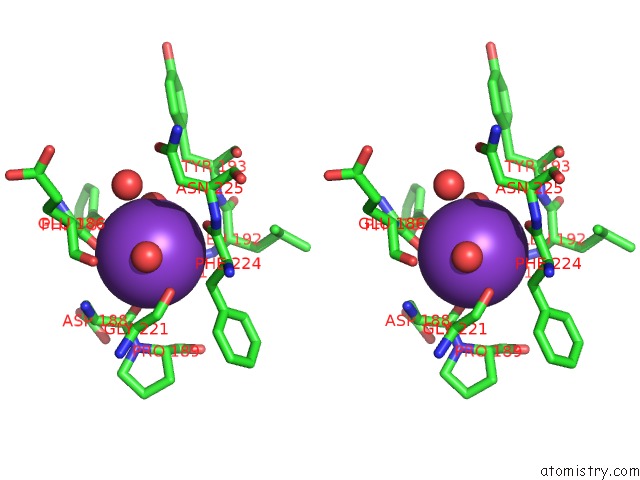
Stereo pair view
Mono view
Stereo pair view
A full contact list of Potassium with other atoms in the K binding
site number 2 of Crystal Structure of Danio Rerio Histone Deacetylase 10 H137A Mutant in Complex with N-Acetylputrescine (Tetrahedral Intermediate) within 5.0Å range:
|
Reference:
C.J.Herbst-Gervasoni,
D.W.Christianson.
X-Ray Crystallographic Snapshots of Substrate Binding in the Active Site of Histone Deacetylase 10. Biochemistry 2021.
ISSN: ISSN 0006-2960
PubMed: 33449614
DOI: 10.1021/ACS.BIOCHEM.0C00936
Page generated: Sat Aug 9 13:33:21 2025
ISSN: ISSN 0006-2960
PubMed: 33449614
DOI: 10.1021/ACS.BIOCHEM.0C00936
Last articles
Mn in 9LJUMn in 9LJW
Mn in 9LJS
Mn in 9LJR
Mn in 9LJT
Mn in 9LJV
Mg in 9UA2
Mg in 9R96
Mg in 9VM1
Mg in 9P01