Potassium »
PDB 6v8y-6w86 »
6v9b »
Potassium in PDB 6v9b: Co-Crystal Structure of the Fluorogenic Mango-IV Homodimer Bound to TO1-Biotin
Protein crystallography data
The structure of Co-Crystal Structure of the Fluorogenic Mango-IV Homodimer Bound to TO1-Biotin, PDB code: 6v9b
was solved by
R.J.Trachman,
A.R.Ferre-D'amare,
with X-Ray Crystallography technique. A brief refinement statistics is given in the table below:
| Resolution Low / High (Å) | 42.39 / 2.40 |
| Space group | P 31 2 1 |
| Cell size a, b, c (Å), α, β, γ (°) | 48.953, 48.953, 123.349, 90.00, 90.00, 120.00 |
| R / Rfree (%) | 20.4 / 24.4 |
Potassium Binding Sites:
The binding sites of Potassium atom in the Co-Crystal Structure of the Fluorogenic Mango-IV Homodimer Bound to TO1-Biotin
(pdb code 6v9b). This binding sites where shown within
5.0 Angstroms radius around Potassium atom.
In total 4 binding sites of Potassium where determined in the Co-Crystal Structure of the Fluorogenic Mango-IV Homodimer Bound to TO1-Biotin, PDB code: 6v9b:
Jump to Potassium binding site number: 1; 2; 3; 4;
In total 4 binding sites of Potassium where determined in the Co-Crystal Structure of the Fluorogenic Mango-IV Homodimer Bound to TO1-Biotin, PDB code: 6v9b:
Jump to Potassium binding site number: 1; 2; 3; 4;
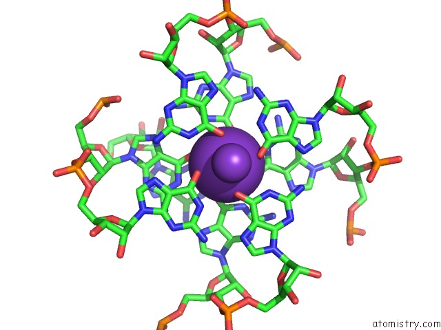
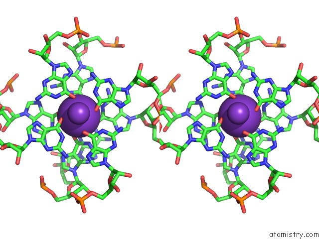
Potassium binding site 1 out of 4 in 6v9b
Go back to
Potassium binding site 1 out
of 4 in the Co-Crystal Structure of the Fluorogenic Mango-IV Homodimer Bound to TO1-Biotin
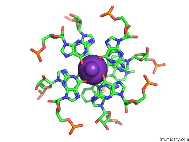
Mono view
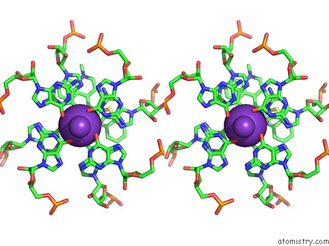
Stereo pair view
Mono view
Stereo pair view
A full contact list of Potassium with other atoms in the K binding
site number 1 of Co-Crystal Structure of the Fluorogenic Mango-IV Homodimer Bound to TO1-Biotin within 5.0Å range:
|
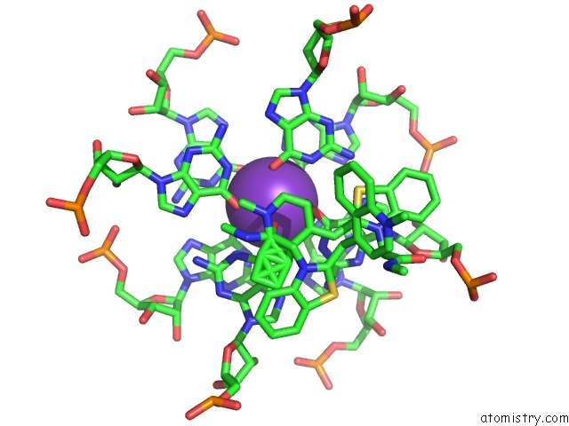
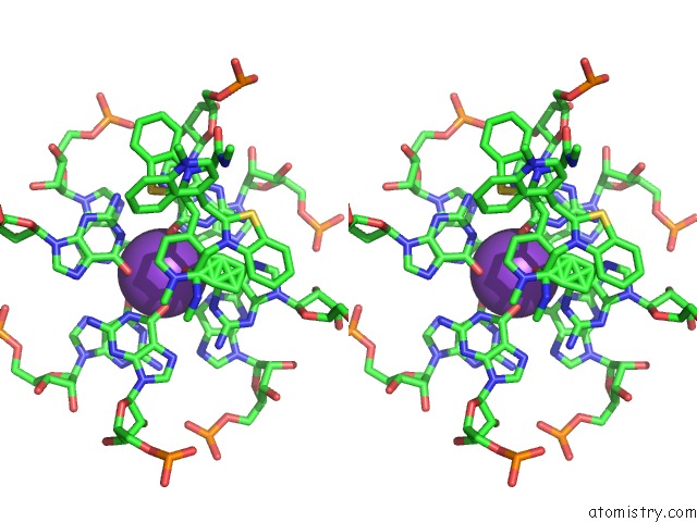
Potassium binding site 2 out of 4 in 6v9b
Go back to
Potassium binding site 2 out
of 4 in the Co-Crystal Structure of the Fluorogenic Mango-IV Homodimer Bound to TO1-Biotin
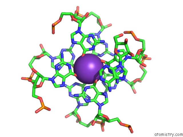
Mono view
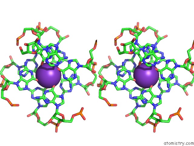
Stereo pair view
Mono view
Stereo pair view
A full contact list of Potassium with other atoms in the K binding
site number 2 of Co-Crystal Structure of the Fluorogenic Mango-IV Homodimer Bound to TO1-Biotin within 5.0Å range:
|
Potassium binding site 3 out of 4 in 6v9b
Go back to
Potassium binding site 3 out
of 4 in the Co-Crystal Structure of the Fluorogenic Mango-IV Homodimer Bound to TO1-Biotin
Mono view
Stereo pair view
Mono view
Stereo pair view
A full contact list of Potassium with other atoms in the K binding
site number 3 of Co-Crystal Structure of the Fluorogenic Mango-IV Homodimer Bound to TO1-Biotin within 5.0Å range:
|
Potassium binding site 4 out of 4 in 6v9b
Go back to
Potassium binding site 4 out
of 4 in the Co-Crystal Structure of the Fluorogenic Mango-IV Homodimer Bound to TO1-Biotin
Mono view
Stereo pair view
Mono view
Stereo pair view
A full contact list of Potassium with other atoms in the K binding
site number 4 of Co-Crystal Structure of the Fluorogenic Mango-IV Homodimer Bound to TO1-Biotin within 5.0Å range:
|
Reference:
R.J.Trachman 3Rd,
R.Cojocaru,
D.Wu,
G.Piszczek,
M.Ryckelynck,
P.J.Unrau,
A.R.Ferre-D'amare.
Structure-Guided Engineering of the Homodimeric Mango-IV Fluorescence Turn-on Aptamer Yields An Rna Fret Pair. Structure 2020.
ISSN: ISSN 0969-2126
PubMed: 32386573
DOI: 10.1016/J.STR.2020.04.007
Page generated: Sat Aug 9 12:26:08 2025
ISSN: ISSN 0969-2126
PubMed: 32386573
DOI: 10.1016/J.STR.2020.04.007
Last articles
Mg in 1VQ4Mg in 1VPA
Mg in 1VPE
Mg in 1VOM
Mg in 1VMA
Mg in 1VMK
Mg in 1VM9
Mg in 1VCR
Mg in 1VLB
Mg in 1VKP