Potassium »
PDB 6u9t-6v7h »
6v36 »
Potassium in PDB 6v36: K2P2.1(Trek-1)I110D Apo Channel Structure
Protein crystallography data
The structure of K2P2.1(Trek-1)I110D Apo Channel Structure, PDB code: 6v36
was solved by
L.Pope,
M.Lolicato,
D.L.Minor,
with X-Ray Crystallography technique. A brief refinement statistics is given in the table below:
| Resolution Low / High (Å) | 14.98 / 3.40 |
| Space group | P 21 21 21 |
| Cell size a, b, c (Å), α, β, γ (°) | 68.192, 120.400, 128.355, 90.00, 90.00, 90.00 |
| R / Rfree (%) | 28.6 / 31.8 |
Other elements in 6v36:
The structure of K2P2.1(Trek-1)I110D Apo Channel Structure also contains other interesting chemical elements:
| Cadmium | (Cd) | 3 atoms |
Potassium Binding Sites:
The binding sites of Potassium atom in the K2P2.1(Trek-1)I110D Apo Channel Structure
(pdb code 6v36). This binding sites where shown within
5.0 Angstroms radius around Potassium atom.
In total 6 binding sites of Potassium where determined in the K2P2.1(Trek-1)I110D Apo Channel Structure, PDB code: 6v36:
Jump to Potassium binding site number: 1; 2; 3; 4; 5; 6;
In total 6 binding sites of Potassium where determined in the K2P2.1(Trek-1)I110D Apo Channel Structure, PDB code: 6v36:
Jump to Potassium binding site number: 1; 2; 3; 4; 5; 6;
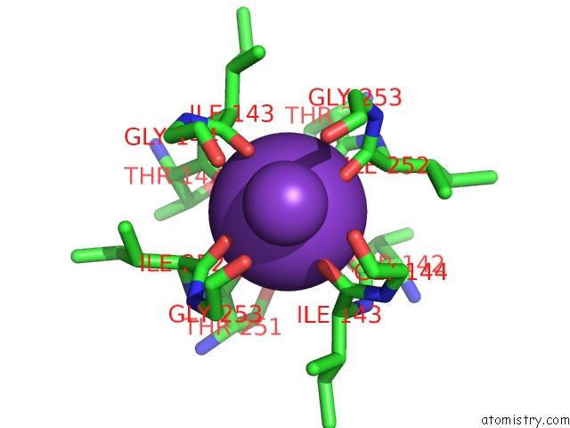
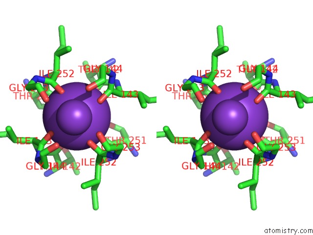
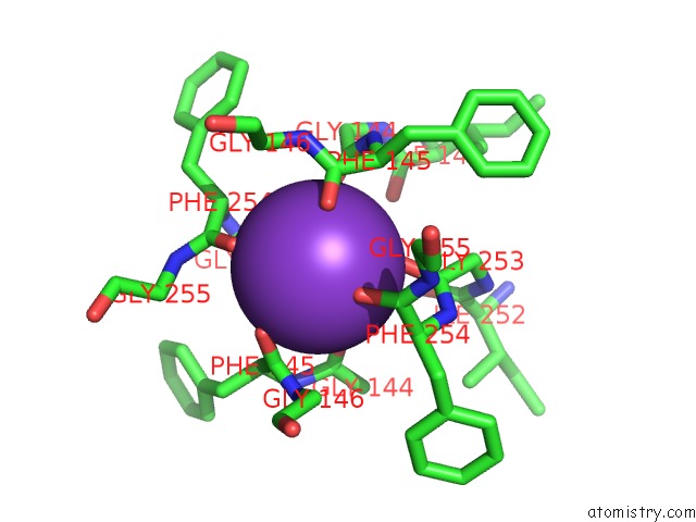
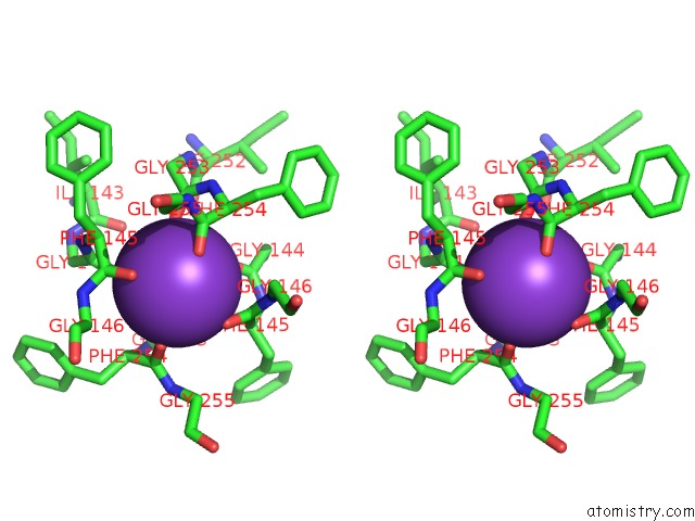
Potassium binding site 1 out of 6 in 6v36
Go back to
Potassium binding site 1 out
of 6 in the K2P2.1(Trek-1)I110D Apo Channel Structure
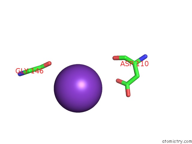
Mono view
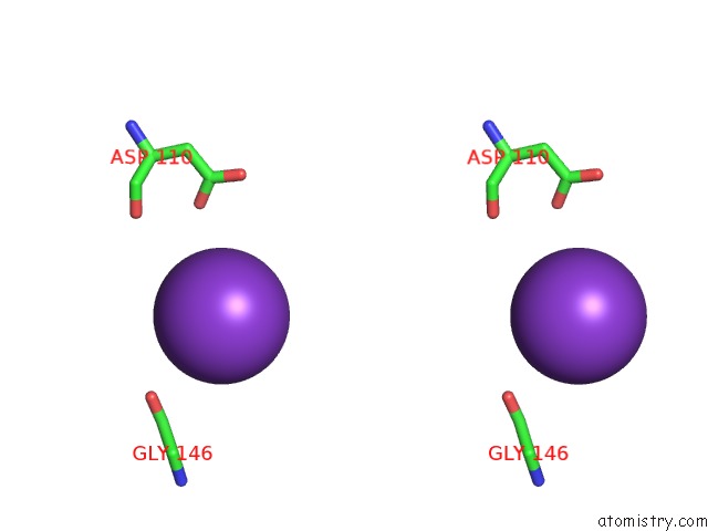
Stereo pair view
Mono view
Stereo pair view
A full contact list of Potassium with other atoms in the K binding
site number 1 of K2P2.1(Trek-1)I110D Apo Channel Structure within 5.0Å range:
|
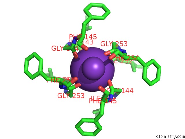
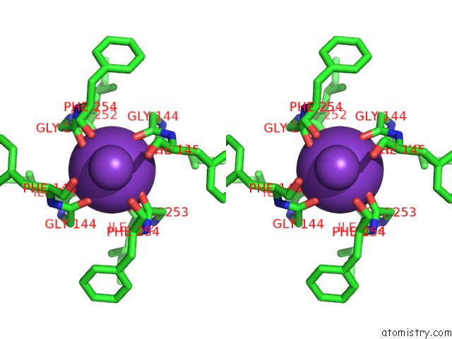
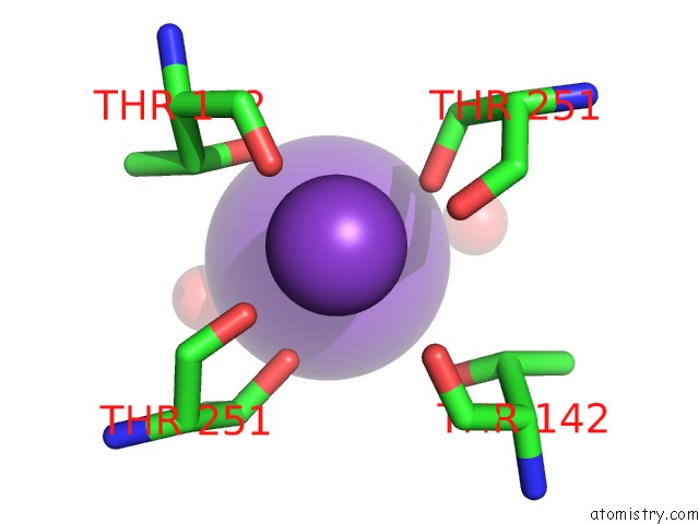
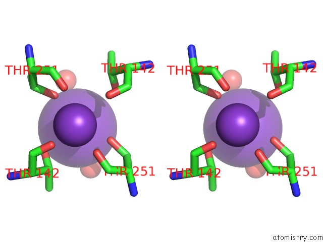
Potassium binding site 2 out of 6 in 6v36
Go back to
Potassium binding site 2 out
of 6 in the K2P2.1(Trek-1)I110D Apo Channel Structure
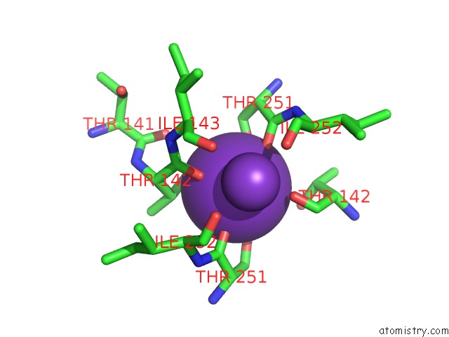
Mono view
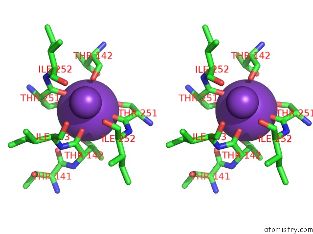
Stereo pair view
Mono view
Stereo pair view
A full contact list of Potassium with other atoms in the K binding
site number 2 of K2P2.1(Trek-1)I110D Apo Channel Structure within 5.0Å range:
|
Potassium binding site 3 out of 6 in 6v36
Go back to
Potassium binding site 3 out
of 6 in the K2P2.1(Trek-1)I110D Apo Channel Structure
Mono view
Stereo pair view
Mono view
Stereo pair view
A full contact list of Potassium with other atoms in the K binding
site number 3 of K2P2.1(Trek-1)I110D Apo Channel Structure within 5.0Å range:
|
Potassium binding site 4 out of 6 in 6v36
Go back to
Potassium binding site 4 out
of 6 in the K2P2.1(Trek-1)I110D Apo Channel Structure
Mono view
Stereo pair view
Mono view
Stereo pair view
A full contact list of Potassium with other atoms in the K binding
site number 4 of K2P2.1(Trek-1)I110D Apo Channel Structure within 5.0Å range:
|
Potassium binding site 5 out of 6 in 6v36
Go back to
Potassium binding site 5 out
of 6 in the K2P2.1(Trek-1)I110D Apo Channel Structure
Mono view
Stereo pair view
Mono view
Stereo pair view
A full contact list of Potassium with other atoms in the K binding
site number 5 of K2P2.1(Trek-1)I110D Apo Channel Structure within 5.0Å range:
|
Potassium binding site 6 out of 6 in 6v36
Go back to
Potassium binding site 6 out
of 6 in the K2P2.1(Trek-1)I110D Apo Channel Structure
Mono view
Stereo pair view
Mono view
Stereo pair view
A full contact list of Potassium with other atoms in the K binding
site number 6 of K2P2.1(Trek-1)I110D Apo Channel Structure within 5.0Å range:
|
Reference:
L.Pope,
M.Lolicato,
D.L.Minor Jr..
Polynuclear Ruthenium Amines Inhibit K2PCHANNELS Via A "Finger in the Dam" Mechanism. Cell Chem Biol 2020.
ISSN: ESSN 2451-9456
PubMed: 32059793
DOI: 10.1016/J.CHEMBIOL.2020.01.011
Page generated: Sat Aug 9 12:22:09 2025
ISSN: ESSN 2451-9456
PubMed: 32059793
DOI: 10.1016/J.CHEMBIOL.2020.01.011
Last articles
Na in 6O6RNa in 6O93
Na in 6O7S
Na in 6O8G
Na in 6O7C
Na in 6O5E
Na in 6O6A
Na in 6O6J
Na in 6O5X
Na in 6NZY