Potassium »
PDB 6pcd-6qm2 »
6pnk »
Potassium in PDB 6pnk: Crystal Structure of the G-Quadruplex Formed By (Gggtt)3GGG in Complex with N-Methylmesoporphryin IX
Protein crystallography data
The structure of Crystal Structure of the G-Quadruplex Formed By (Gggtt)3GGG in Complex with N-Methylmesoporphryin IX, PDB code: 6pnk
was solved by
L.A.Yatsunyk,
L.Y.Lin,
with X-Ray Crystallography technique. A brief refinement statistics is given in the table below:
| Resolution Low / High (Å) | 64.78 / 2.39 |
| Space group | H 3 2 |
| Cell size a, b, c (Å), α, β, γ (°) | 60.930, 60.930, 194.342, 90.00, 90.00, 120.00 |
| R / Rfree (%) | 23.5 / 24.9 |
Other elements in 6pnk:
The structure of Crystal Structure of the G-Quadruplex Formed By (Gggtt)3GGG in Complex with N-Methylmesoporphryin IX also contains other interesting chemical elements:
| Sodium | (Na) | 1 atom |
Potassium Binding Sites:
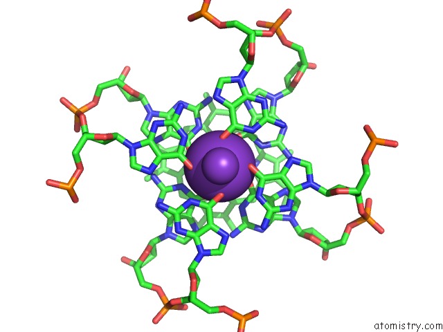
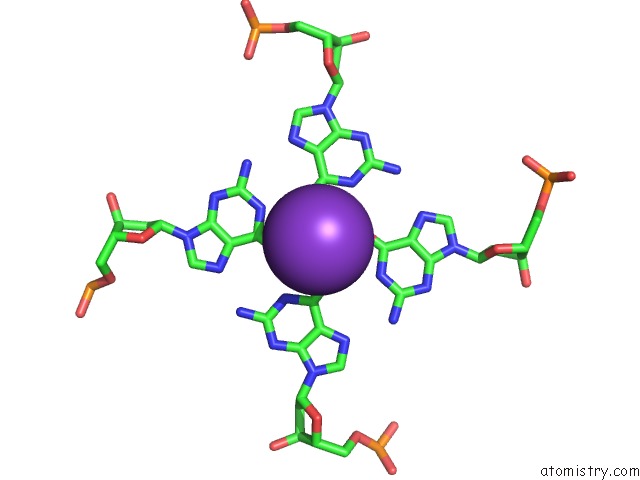
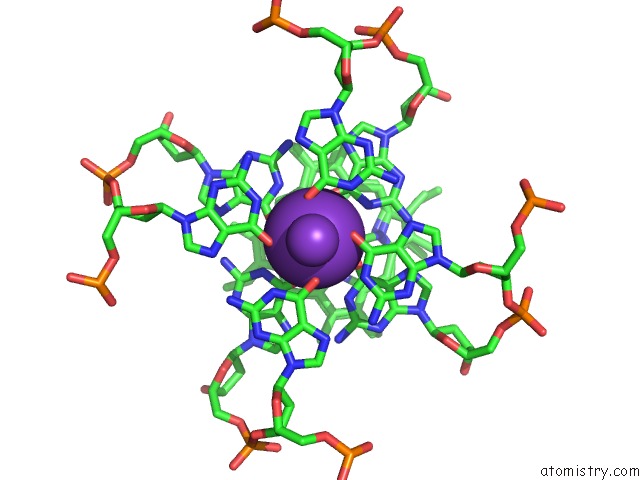
The binding sites of Potassium atom in the Crystal Structure of the G-Quadruplex Formed By (Gggtt)3GGG in Complex with N-Methylmesoporphryin IX
(pdb code 6pnk). This binding sites where shown within
5.0 Angstroms radius around Potassium atom.
In total 6 binding sites of Potassium where determined in the Crystal Structure of the G-Quadruplex Formed By (Gggtt)3GGG in Complex with N-Methylmesoporphryin IX, PDB code: 6pnk:
Jump to Potassium binding site number: 1; 2; 3; 4; 5; 6;
In total 6 binding sites of Potassium where determined in the Crystal Structure of the G-Quadruplex Formed By (Gggtt)3GGG in Complex with N-Methylmesoporphryin IX, PDB code: 6pnk:
Jump to Potassium binding site number: 1; 2; 3; 4; 5; 6;
Potassium binding site 1 out of 6 in 6pnk
Go back to
Potassium binding site 1 out
of 6 in the Crystal Structure of the G-Quadruplex Formed By (Gggtt)3GGG in Complex with N-Methylmesoporphryin IX

Mono view

Stereo pair view
Mono view
Stereo pair view
A full contact list of Potassium with other atoms in the K binding
site number 1 of Crystal Structure of the G-Quadruplex Formed By (Gggtt)3GGG in Complex with N-Methylmesoporphryin IX within 5.0Å range:
|
Potassium binding site 2 out of 6 in 6pnk
Go back to
Potassium binding site 2 out
of 6 in the Crystal Structure of the G-Quadruplex Formed By (Gggtt)3GGG in Complex with N-Methylmesoporphryin IX
Mono view

Stereo pair view
Mono view
Stereo pair view
A full contact list of Potassium with other atoms in the K binding
site number 2 of Crystal Structure of the G-Quadruplex Formed By (Gggtt)3GGG in Complex with N-Methylmesoporphryin IX within 5.0Å range:
|
Potassium binding site 3 out of 6 in 6pnk
Go back to
Potassium binding site 3 out
of 6 in the Crystal Structure of the G-Quadruplex Formed By (Gggtt)3GGG in Complex with N-Methylmesoporphryin IX
Mono view

Stereo pair view
Mono view
Stereo pair view
A full contact list of Potassium with other atoms in the K binding
site number 3 of Crystal Structure of the G-Quadruplex Formed By (Gggtt)3GGG in Complex with N-Methylmesoporphryin IX within 5.0Å range:
|
Potassium binding site 4 out of 6 in 6pnk
Go back to
Potassium binding site 4 out
of 6 in the Crystal Structure of the G-Quadruplex Formed By (Gggtt)3GGG in Complex with N-Methylmesoporphryin IX
Mono view

Stereo pair view
Mono view
Stereo pair view
A full contact list of Potassium with other atoms in the K binding
site number 4 of Crystal Structure of the G-Quadruplex Formed By (Gggtt)3GGG in Complex with N-Methylmesoporphryin IX within 5.0Å range:
|
Potassium binding site 5 out of 6 in 6pnk
Go back to
Potassium binding site 5 out
of 6 in the Crystal Structure of the G-Quadruplex Formed By (Gggtt)3GGG in Complex with N-Methylmesoporphryin IX

Mono view

Stereo pair view
Mono view
Stereo pair view
A full contact list of Potassium with other atoms in the K binding
site number 5 of Crystal Structure of the G-Quadruplex Formed By (Gggtt)3GGG in Complex with N-Methylmesoporphryin IX within 5.0Å range:
|
Potassium binding site 6 out of 6 in 6pnk
Go back to
Potassium binding site 6 out
of 6 in the Crystal Structure of the G-Quadruplex Formed By (Gggtt)3GGG in Complex with N-Methylmesoporphryin IX

Mono view

Stereo pair view
Mono view
Stereo pair view
A full contact list of Potassium with other atoms in the K binding
site number 6 of Crystal Structure of the G-Quadruplex Formed By (Gggtt)3GGG in Complex with N-Methylmesoporphryin IX within 5.0Å range:
|
Reference:
L.Y.Lin,
S.Mccarthy,
B.M.Powell,
Y.Manurung,
I.M.Xiang,
W.L.Dean,
B.Chaires,
L.A.Yatsunyk.
Biophysical and X-Ray Structural Studies of the (Gggtt)3GGG G-Quadruplex in Complex with N-Methyl Mesoporphyrin IX. Plos One V. 15 41513 2020.
ISSN: ESSN 1932-6203
PubMed: 33206666
DOI: 10.1371/JOURNAL.PONE.0241513
Page generated: Mon Aug 12 17:12:40 2024
ISSN: ESSN 1932-6203
PubMed: 33206666
DOI: 10.1371/JOURNAL.PONE.0241513
Last articles
I in 4R11I in 4RCE
I in 4RCD
I in 4R1T
I in 4QRP
I in 4QU3
I in 4PXS
I in 4QJQ
I in 4Q53
I in 4Q2M