Potassium »
PDB 6ldm-6n92 »
6mgu »
Potassium in PDB 6mgu: Crystal Structure of the Catalytic Domain of the Inosine Monophosphate Dehydrogenase From Bacillus Anthracis in the Complex with Inhibitor Oxanosine Monophosphate
Enzymatic activity of Crystal Structure of the Catalytic Domain of the Inosine Monophosphate Dehydrogenase From Bacillus Anthracis in the Complex with Inhibitor Oxanosine Monophosphate
All present enzymatic activity of Crystal Structure of the Catalytic Domain of the Inosine Monophosphate Dehydrogenase From Bacillus Anthracis in the Complex with Inhibitor Oxanosine Monophosphate:
1.1.1.205;
1.1.1.205;
Protein crystallography data
The structure of Crystal Structure of the Catalytic Domain of the Inosine Monophosphate Dehydrogenase From Bacillus Anthracis in the Complex with Inhibitor Oxanosine Monophosphate, PDB code: 6mgu
was solved by
Y.Kim,
N.Maltseva,
R.Yu,
L.Hedstrom,
A.Joachimiak,
Center For Structuralgenomics Of Infectious Diseases (Csgid),
with X-Ray Crystallography technique. A brief refinement statistics is given in the table below:
| Resolution Low / High (Å) | 43.21 / 1.54 |
| Space group | P 4 |
| Cell size a, b, c (Å), α, β, γ (°) | 86.411, 86.411, 90.984, 90.00, 90.00, 90.00 |
| R / Rfree (%) | 16.5 / 19.8 |
Potassium Binding Sites:
The binding sites of Potassium atom in the Crystal Structure of the Catalytic Domain of the Inosine Monophosphate Dehydrogenase From Bacillus Anthracis in the Complex with Inhibitor Oxanosine Monophosphate
(pdb code 6mgu). This binding sites where shown within
5.0 Angstroms radius around Potassium atom.
In total 2 binding sites of Potassium where determined in the Crystal Structure of the Catalytic Domain of the Inosine Monophosphate Dehydrogenase From Bacillus Anthracis in the Complex with Inhibitor Oxanosine Monophosphate, PDB code: 6mgu:
Jump to Potassium binding site number: 1; 2;
In total 2 binding sites of Potassium where determined in the Crystal Structure of the Catalytic Domain of the Inosine Monophosphate Dehydrogenase From Bacillus Anthracis in the Complex with Inhibitor Oxanosine Monophosphate, PDB code: 6mgu:
Jump to Potassium binding site number: 1; 2;
Potassium binding site 1 out of 2 in 6mgu
Go back to
Potassium binding site 1 out
of 2 in the Crystal Structure of the Catalytic Domain of the Inosine Monophosphate Dehydrogenase From Bacillus Anthracis in the Complex with Inhibitor Oxanosine Monophosphate
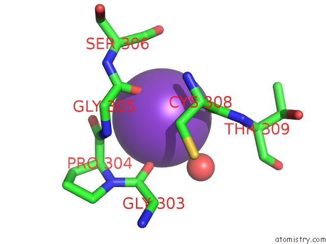
Mono view
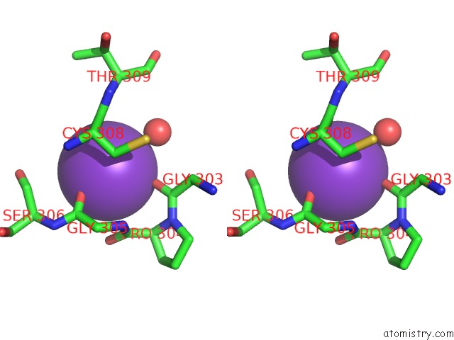
Stereo pair view
Mono view
Stereo pair view
A full contact list of Potassium with other atoms in the K binding
site number 1 of Crystal Structure of the Catalytic Domain of the Inosine Monophosphate Dehydrogenase From Bacillus Anthracis in the Complex with Inhibitor Oxanosine Monophosphate within 5.0Å range:
|
Potassium binding site 2 out of 2 in 6mgu
Go back to
Potassium binding site 2 out
of 2 in the Crystal Structure of the Catalytic Domain of the Inosine Monophosphate Dehydrogenase From Bacillus Anthracis in the Complex with Inhibitor Oxanosine Monophosphate
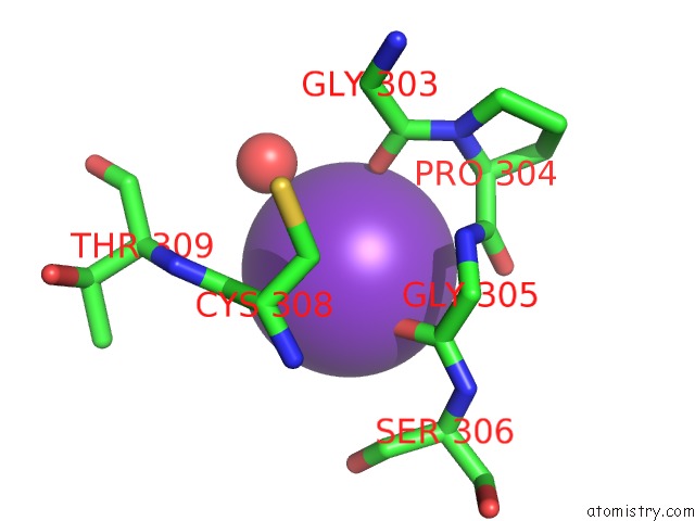
Mono view
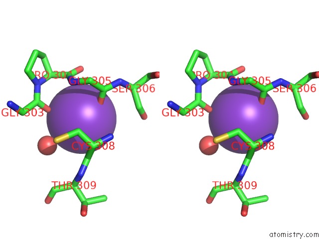
Stereo pair view
Mono view
Stereo pair view
A full contact list of Potassium with other atoms in the K binding
site number 2 of Crystal Structure of the Catalytic Domain of the Inosine Monophosphate Dehydrogenase From Bacillus Anthracis in the Complex with Inhibitor Oxanosine Monophosphate within 5.0Å range:
|
Reference:
Y.Kim,
N.Maltseva,
R.Yu,
L.Hedstrom,
A.Joachimiak,
Center For Structural Genomics Of Infectious Diseases(Csgid).
Crystal Structure of the Catalytic Domain of the Inosine Monophosphate Dehydrogenase From Bacillus Anthracis in the Complex with Inhibitor Oxanosine Monophosphate To Be Published.
Page generated: Sat Aug 9 11:33:54 2025
Last articles
La in 5YTQLa in 6IP9
La in 1DJG
La in 6DAM
La in 5KKB
La in 2OQR
La in 2RPV
La in 5KIJ
La in 2K0J
La in 2I18