Potassium »
PDB 6i4i-6lab »
6ky3 »
Potassium in PDB 6ky3: Structure of Arginine Kinase H284A Mutant
Protein crystallography data
The structure of Structure of Arginine Kinase H284A Mutant, PDB code: 6ky3
was solved by
Z.Rao,
J.H.Park,
S.Y.Kim,
D.S.Kim,
with X-Ray Crystallography technique. A brief refinement statistics is given in the table below:
| Resolution Low / High (Å) | 46.17 / 1.34 |
| Space group | C 1 2 1 |
| Cell size a, b, c (Å), α, β, γ (°) | 77.943, 57.840, 74.860, 90.00, 100.46, 90.00 |
| R / Rfree (%) | 17.3 / 21.8 |
Potassium Binding Sites:
The binding sites of Potassium atom in the Structure of Arginine Kinase H284A Mutant
(pdb code 6ky3). This binding sites where shown within
5.0 Angstroms radius around Potassium atom.
In total 2 binding sites of Potassium where determined in the Structure of Arginine Kinase H284A Mutant, PDB code: 6ky3:
Jump to Potassium binding site number: 1; 2;
In total 2 binding sites of Potassium where determined in the Structure of Arginine Kinase H284A Mutant, PDB code: 6ky3:
Jump to Potassium binding site number: 1; 2;
Potassium binding site 1 out of 2 in 6ky3
Go back to
Potassium binding site 1 out
of 2 in the Structure of Arginine Kinase H284A Mutant
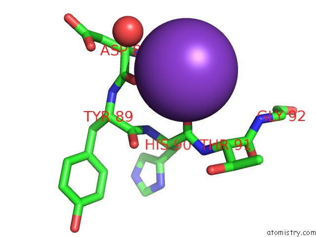
Mono view
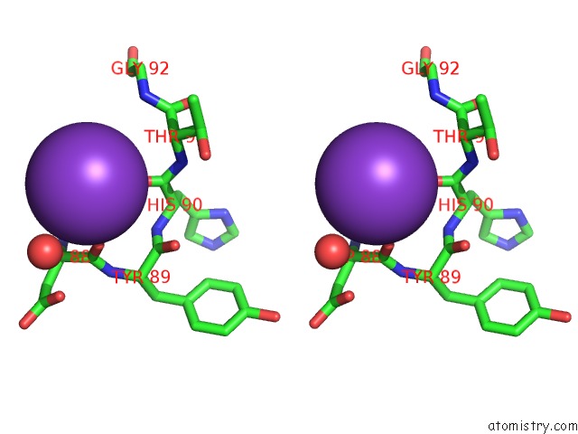
Stereo pair view
Mono view
Stereo pair view
A full contact list of Potassium with other atoms in the K binding
site number 1 of Structure of Arginine Kinase H284A Mutant within 5.0Å range:
|
Potassium binding site 2 out of 2 in 6ky3
Go back to
Potassium binding site 2 out
of 2 in the Structure of Arginine Kinase H284A Mutant
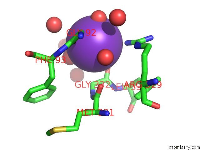
Mono view
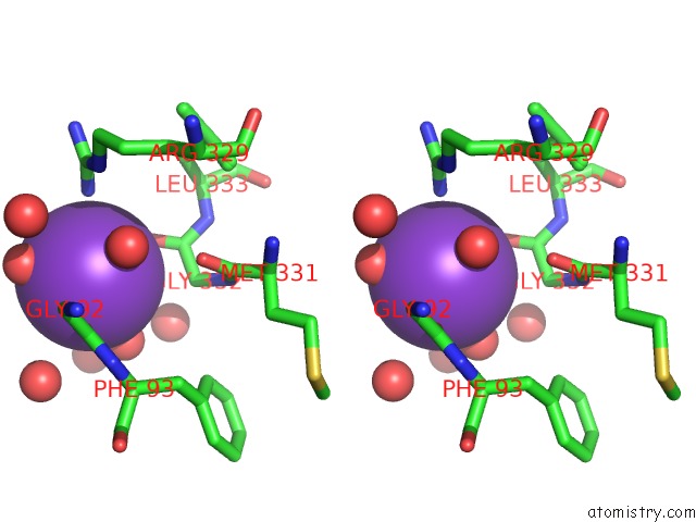
Stereo pair view
Mono view
Stereo pair view
A full contact list of Potassium with other atoms in the K binding
site number 2 of Structure of Arginine Kinase H284A Mutant within 5.0Å range:
|
Reference:
Z.Rao,
J.H.Park,
S.Y.Kim,
D.S.Kim.
Structure of Arginine Kinase H284A Mutant To Be Published.
Page generated: Sat Aug 9 11:30:03 2025
Last articles
Mo in 3K7RMo in 3ML1
Mo in 3K6X
Mo in 3L4P
Mo in 3K1A
Mo in 3K6W
Mo in 3GZG
Mo in 3HRD
Mo in 3IR5
Mo in 3IR7