Potassium »
PDB 5vt3-5x23 »
5wnn »
Potassium in PDB 5wnn: Crystal Structure of Phosphate-Binding Protein Psts Protein From Burkholderia Pseudomallei
Protein crystallography data
The structure of Crystal Structure of Phosphate-Binding Protein Psts Protein From Burkholderia Pseudomallei, PDB code: 5wnn
was solved by
Seattle Structural Genomics Center For Infectious Disease (Ssgcid),
with X-Ray Crystallography technique. A brief refinement statistics is given in the table below:
| Resolution Low / High (Å) | 43.34 / 1.85 |
| Space group | C 1 2 1 |
| Cell size a, b, c (Å), α, β, γ (°) | 116.640, 59.840, 101.090, 90.00, 120.97, 90.00 |
| R / Rfree (%) | 17 / 21.8 |
Potassium Binding Sites:
The binding sites of Potassium atom in the Crystal Structure of Phosphate-Binding Protein Psts Protein From Burkholderia Pseudomallei
(pdb code 5wnn). This binding sites where shown within
5.0 Angstroms radius around Potassium atom.
In total 2 binding sites of Potassium where determined in the Crystal Structure of Phosphate-Binding Protein Psts Protein From Burkholderia Pseudomallei, PDB code: 5wnn:
Jump to Potassium binding site number: 1; 2;
In total 2 binding sites of Potassium where determined in the Crystal Structure of Phosphate-Binding Protein Psts Protein From Burkholderia Pseudomallei, PDB code: 5wnn:
Jump to Potassium binding site number: 1; 2;
Potassium binding site 1 out of 2 in 5wnn
Go back to
Potassium binding site 1 out
of 2 in the Crystal Structure of Phosphate-Binding Protein Psts Protein From Burkholderia Pseudomallei
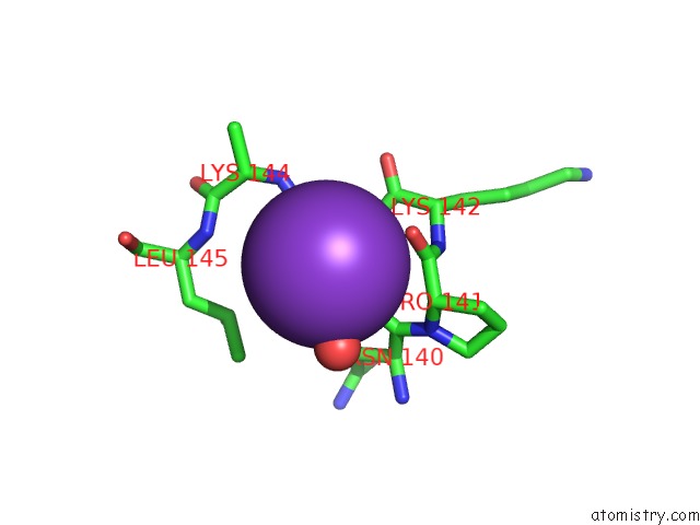
Mono view
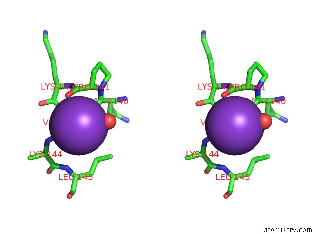
Stereo pair view
Mono view
Stereo pair view
A full contact list of Potassium with other atoms in the K binding
site number 1 of Crystal Structure of Phosphate-Binding Protein Psts Protein From Burkholderia Pseudomallei within 5.0Å range:
|
Potassium binding site 2 out of 2 in 5wnn
Go back to
Potassium binding site 2 out
of 2 in the Crystal Structure of Phosphate-Binding Protein Psts Protein From Burkholderia Pseudomallei
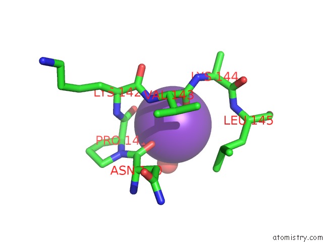
Mono view
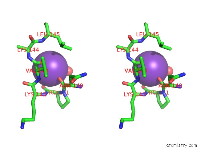
Stereo pair view
Mono view
Stereo pair view
A full contact list of Potassium with other atoms in the K binding
site number 2 of Crystal Structure of Phosphate-Binding Protein Psts Protein From Burkholderia Pseudomallei within 5.0Å range:
|
Reference:
J.Abendroth,
D.M.Dranow,
D.D.Lorimer,
T.E.Edwards.
Crystal Structure of Phosphate-Binding Protein Psts Protein From Burkholderia Pseudomallei To Be Published.
Page generated: Sat Aug 9 10:06:29 2025
Last articles
Na in 2R22Na in 2R20
Na in 2R21
Na in 2QZ7
Na in 2QZI
Na in 2R1S
Na in 2R0W
Na in 2QXF
Na in 2QZ2
Na in 2QWP