Potassium »
PDB 5u3s-5vsn »
5ua3 »
Potassium in PDB 5ua3: Crystal Structure of A Dna G-Quadruplex with A Cytosine Bulge
Protein crystallography data
The structure of Crystal Structure of A Dna G-Quadruplex with A Cytosine Bulge, PDB code: 5ua3
was solved by
M.Meier,
M.D.Mcdougall,
N.J.Krahn,
A.Moya-Torres,
E.K.S.Mcrae,
E.P.Booy,
T.R.Patel,
S.A.Mckenna,
J.Stetefeld,
with X-Ray Crystallography technique. A brief refinement statistics is given in the table below:
| Resolution Low / High (Å) | 19.49 / 1.88 |
| Space group | P 1 |
| Cell size a, b, c (Å), α, β, γ (°) | 30.103, 33.100, 33.282, 64.65, 78.64, 81.78 |
| R / Rfree (%) | 22.7 / 24.7 |
Potassium Binding Sites:
The binding sites of Potassium atom in the Crystal Structure of A Dna G-Quadruplex with A Cytosine Bulge
(pdb code 5ua3). This binding sites where shown within
5.0 Angstroms radius around Potassium atom.
In total 5 binding sites of Potassium where determined in the Crystal Structure of A Dna G-Quadruplex with A Cytosine Bulge, PDB code: 5ua3:
Jump to Potassium binding site number: 1; 2; 3; 4; 5;
In total 5 binding sites of Potassium where determined in the Crystal Structure of A Dna G-Quadruplex with A Cytosine Bulge, PDB code: 5ua3:
Jump to Potassium binding site number: 1; 2; 3; 4; 5;
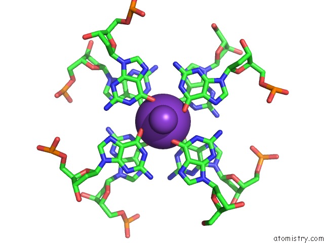
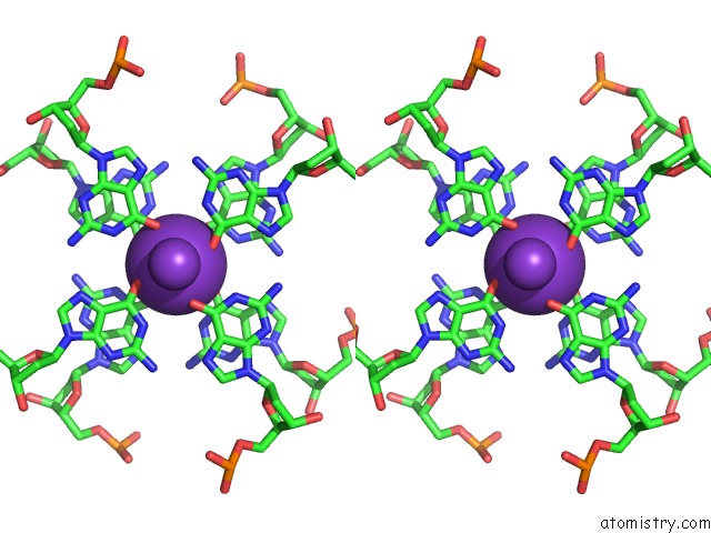
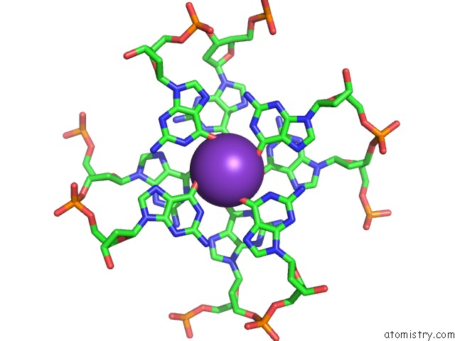
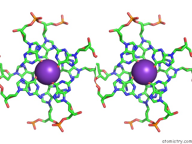
Potassium binding site 1 out of 5 in 5ua3
Go back to
Potassium binding site 1 out
of 5 in the Crystal Structure of A Dna G-Quadruplex with A Cytosine Bulge
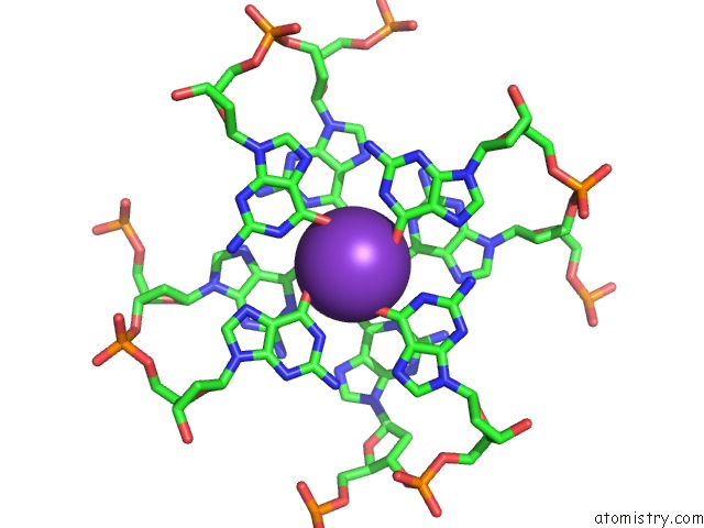
Mono view
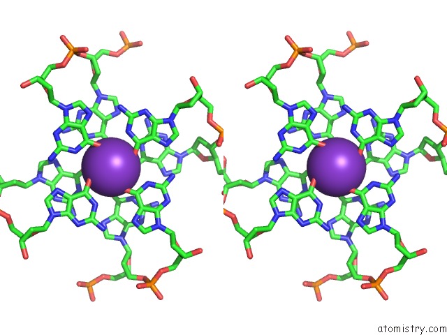
Stereo pair view
Mono view
Stereo pair view
A full contact list of Potassium with other atoms in the K binding
site number 1 of Crystal Structure of A Dna G-Quadruplex with A Cytosine Bulge within 5.0Å range:
|
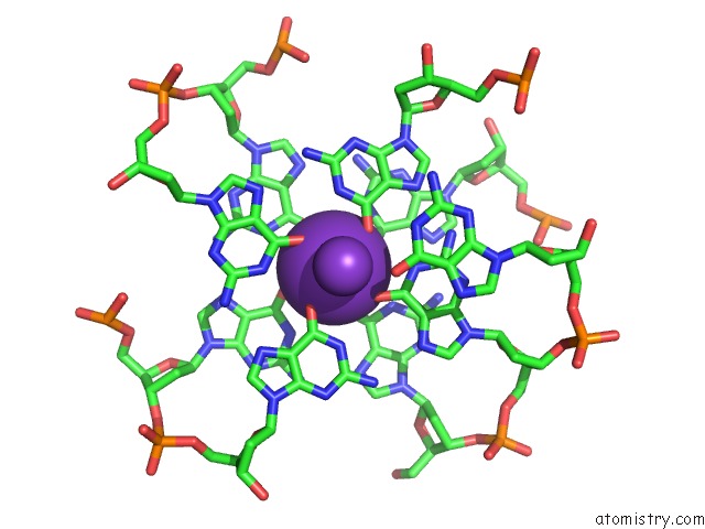
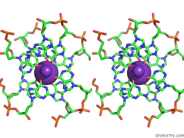
Potassium binding site 2 out of 5 in 5ua3
Go back to
Potassium binding site 2 out
of 5 in the Crystal Structure of A Dna G-Quadruplex with A Cytosine Bulge
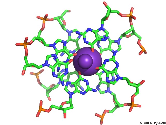
Mono view
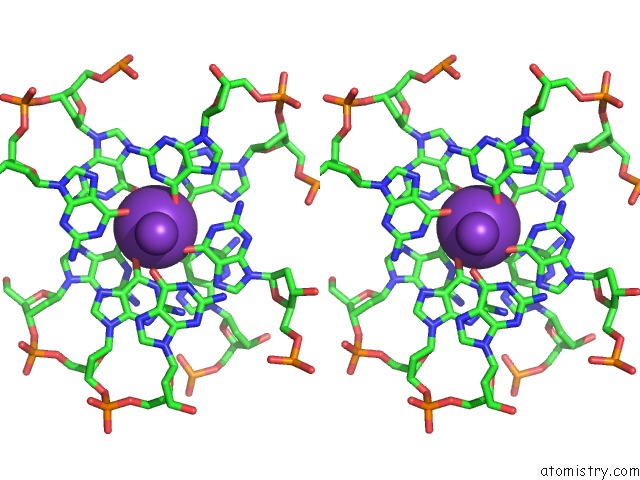
Stereo pair view
Mono view
Stereo pair view
A full contact list of Potassium with other atoms in the K binding
site number 2 of Crystal Structure of A Dna G-Quadruplex with A Cytosine Bulge within 5.0Å range:
|
Potassium binding site 3 out of 5 in 5ua3
Go back to
Potassium binding site 3 out
of 5 in the Crystal Structure of A Dna G-Quadruplex with A Cytosine Bulge
Mono view
Stereo pair view
Mono view
Stereo pair view
A full contact list of Potassium with other atoms in the K binding
site number 3 of Crystal Structure of A Dna G-Quadruplex with A Cytosine Bulge within 5.0Å range:
|
Potassium binding site 4 out of 5 in 5ua3
Go back to
Potassium binding site 4 out
of 5 in the Crystal Structure of A Dna G-Quadruplex with A Cytosine Bulge
Mono view
Stereo pair view
Mono view
Stereo pair view
A full contact list of Potassium with other atoms in the K binding
site number 4 of Crystal Structure of A Dna G-Quadruplex with A Cytosine Bulge within 5.0Å range:
|
Potassium binding site 5 out of 5 in 5ua3
Go back to
Potassium binding site 5 out
of 5 in the Crystal Structure of A Dna G-Quadruplex with A Cytosine Bulge
Mono view
Stereo pair view
Mono view
Stereo pair view
A full contact list of Potassium with other atoms in the K binding
site number 5 of Crystal Structure of A Dna G-Quadruplex with A Cytosine Bulge within 5.0Å range:
|
Reference:
M.Meier,
A.Moya-Torres,
N.J.Krahn,
M.D.Mcdougall,
G.L.Orriss,
E.K.S.Mcrae,
E.P.Booy,
K.Mceleney,
T.R.Patel,
S.A.Mckenna,
J.Stetefeld.
Structure and Hydrodynamics of A Dna G-Quadruplex with A Cytosine Bulge. Nucleic Acids Res. V. 46 5319 2018.
ISSN: ESSN 1362-4962
PubMed: 29718405
DOI: 10.1093/NAR/GKY307
Page generated: Sat Aug 9 09:51:59 2025
ISSN: ESSN 1362-4962
PubMed: 29718405
DOI: 10.1093/NAR/GKY307
Last articles
Na in 3HWFNa in 3HWE
Na in 3HWT
Na in 3HWD
Na in 3HW8
Na in 3HVY
Na in 3HVU
Na in 3HHQ
Na in 3HVI
Na in 3HSS