Potassium »
PDB 5sc8-5u3q »
5txg »
Potassium in PDB 5txg: Crystal Structure of the Zika Virus NS3 Helicase.
Protein crystallography data
The structure of Crystal Structure of the Zika Virus NS3 Helicase., PDB code: 5txg
was solved by
S.Nocadello,
S.H.Light,
G.Minasov,
L.Shuvalova,
A.A.Cardona-Correa,
I.Ojeda,
J.Vargas,
M.E.Johnson,
H.Lee,
W.F.Anderson,
Center Forstructural Genomics Of Infectious Diseases (Csgid),
with X-Ray Crystallography technique. A brief refinement statistics is given in the table below:
| Resolution Low / High (Å) | 29.97 / 2.05 |
| Space group | P 1 21 1 |
| Cell size a, b, c (Å), α, β, γ (°) | 51.639, 69.855, 58.461, 90.00, 93.41, 90.00 |
| R / Rfree (%) | 18.4 / 24.7 |
Potassium Binding Sites:
The binding sites of Potassium atom in the Crystal Structure of the Zika Virus NS3 Helicase.
(pdb code 5txg). This binding sites where shown within
5.0 Angstroms radius around Potassium atom.
In total 2 binding sites of Potassium where determined in the Crystal Structure of the Zika Virus NS3 Helicase., PDB code: 5txg:
Jump to Potassium binding site number: 1; 2;
In total 2 binding sites of Potassium where determined in the Crystal Structure of the Zika Virus NS3 Helicase., PDB code: 5txg:
Jump to Potassium binding site number: 1; 2;
Potassium binding site 1 out of 2 in 5txg
Go back to
Potassium binding site 1 out
of 2 in the Crystal Structure of the Zika Virus NS3 Helicase.
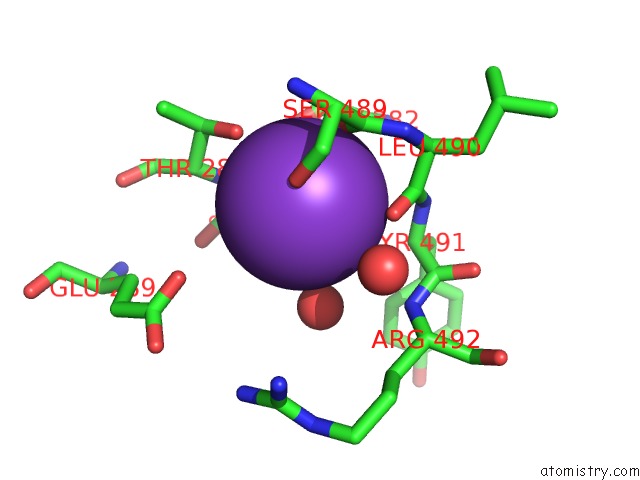
Mono view
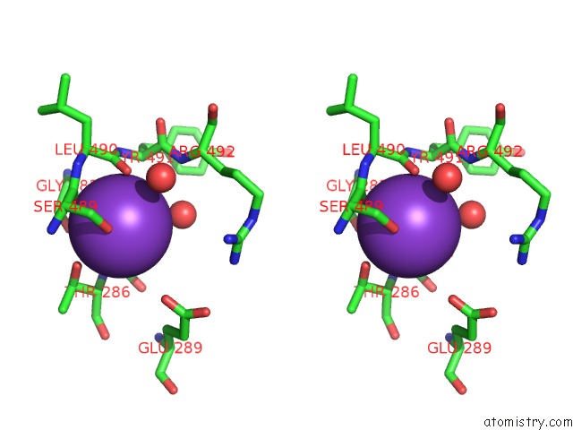
Stereo pair view
Mono view
Stereo pair view
A full contact list of Potassium with other atoms in the K binding
site number 1 of Crystal Structure of the Zika Virus NS3 Helicase. within 5.0Å range:
|
Potassium binding site 2 out of 2 in 5txg
Go back to
Potassium binding site 2 out
of 2 in the Crystal Structure of the Zika Virus NS3 Helicase.
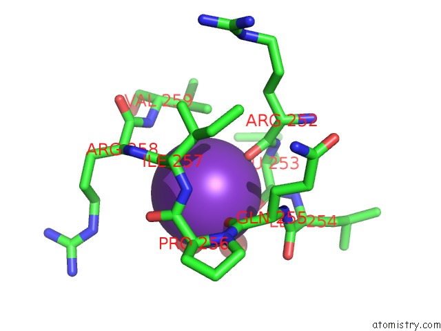
Mono view
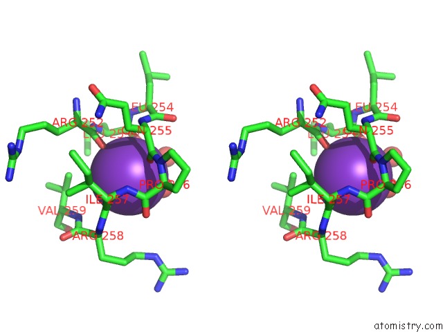
Stereo pair view
Mono view
Stereo pair view
A full contact list of Potassium with other atoms in the K binding
site number 2 of Crystal Structure of the Zika Virus NS3 Helicase. within 5.0Å range:
|
Reference:
S.Nocadello,
S.H.Light,
G.Minasov,
L.Shuvalova,
A.A.Cardona-Correa,
I.Ojeda,
J.Vargas,
M.E.Johnson,
H.Lee,
W.F.Anderson,
Center For Structural Genomics Of Infectious Diseases(Csgid).
Crystal Structure of the Zika Virus NS3 Helicase. To Be Published.
Page generated: Sat Aug 9 09:51:13 2025
Last articles
Mg in 6HNQMg in 6HOS
Mg in 6HNS
Mg in 6HN2
Mg in 6HMZ
Mg in 6HMU
Mg in 6HMT
Mg in 6HLR
Mg in 6HLQ
Mg in 6HKY