Potassium »
PDB 5ksd-5mq0 »
5ls8 »
Potassium in PDB 5ls8: Light-Activated Ruthenium Complex Bound to A Dna Quadruplex
Protein crystallography data
The structure of Light-Activated Ruthenium Complex Bound to A Dna Quadruplex, PDB code: 5ls8
was solved by
K.T.Mcquaid,
H.Abell,
J.P.Hall,
C.J.Cardin,
with X-Ray Crystallography technique. A brief refinement statistics is given in the table below:
| Resolution Low / High (Å) | 31.72 / 1.78 |
| Space group | P 65 |
| Cell size a, b, c (Å), α, β, γ (°) | 36.625, 36.625, 136.104, 90.00, 90.00, 120.00 |
| R / Rfree (%) | 21.4 / 23.3 |
Other elements in 5ls8:
The structure of Light-Activated Ruthenium Complex Bound to A Dna Quadruplex also contains other interesting chemical elements:
| Ruthenium | (Ru) | 6 atoms |
Potassium Binding Sites:
The binding sites of Potassium atom in the Light-Activated Ruthenium Complex Bound to A Dna Quadruplex
(pdb code 5ls8). This binding sites where shown within
5.0 Angstroms radius around Potassium atom.
In total 2 binding sites of Potassium where determined in the Light-Activated Ruthenium Complex Bound to A Dna Quadruplex, PDB code: 5ls8:
Jump to Potassium binding site number: 1; 2;
In total 2 binding sites of Potassium where determined in the Light-Activated Ruthenium Complex Bound to A Dna Quadruplex, PDB code: 5ls8:
Jump to Potassium binding site number: 1; 2;
Potassium binding site 1 out of 2 in 5ls8
Go back to
Potassium binding site 1 out
of 2 in the Light-Activated Ruthenium Complex Bound to A Dna Quadruplex
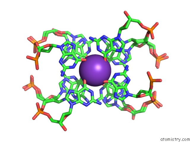
Mono view
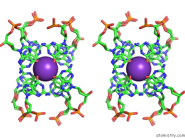
Stereo pair view
Mono view
Stereo pair view
A full contact list of Potassium with other atoms in the K binding
site number 1 of Light-Activated Ruthenium Complex Bound to A Dna Quadruplex within 5.0Å range:
|
Potassium binding site 2 out of 2 in 5ls8
Go back to
Potassium binding site 2 out
of 2 in the Light-Activated Ruthenium Complex Bound to A Dna Quadruplex
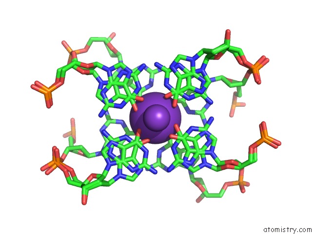
Mono view
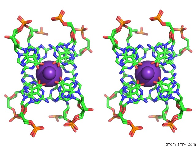
Stereo pair view
Mono view
Stereo pair view
A full contact list of Potassium with other atoms in the K binding
site number 2 of Light-Activated Ruthenium Complex Bound to A Dna Quadruplex within 5.0Å range:
|
Reference:
K.Mcquaid,
H.Abell,
S.P.Gurung,
D.R.Allan,
G.Winter,
T.Sorensen,
D.J.Cardin,
J.A.Brazier,
C.J.Cardin,
J.P.Hall.
Structural Studies Reveal Enantiospecific Recognition of A Dna G-Quadruplex By A Ruthenium Polypyridyl Complex. Angew.Chem.Int.Ed.Engl. V. 58 9881 2019.
ISSN: ESSN 1521-3773
PubMed: 30958918
DOI: 10.1002/ANIE.201814502
Page generated: Sat Aug 9 09:32:19 2025
ISSN: ESSN 1521-3773
PubMed: 30958918
DOI: 10.1002/ANIE.201814502
Last articles
Mn in 7DYWMn in 7D7Z
Mn in 7DYV
Mn in 7DYU
Mn in 7DYT
Mn in 7DNQ
Mn in 7DNN
Mn in 7DDW
Mn in 7DNM
Mn in 7DMM