Potassium »
PDB 5g17-5imu »
5hv4 »
Potassium in PDB 5hv4: Crystal Structure of A Prolyl 4-Hydroxylase Complexed with Alpha- Ketoglutarate From the Pathogenic Bacterium Bacillus Anthracis in C2221
Protein crystallography data
The structure of Crystal Structure of A Prolyl 4-Hydroxylase Complexed with Alpha- Ketoglutarate From the Pathogenic Bacterium Bacillus Anthracis in C2221, PDB code: 5hv4
was solved by
N.J.Schnicker,
M.Dey,
with X-Ray Crystallography technique. A brief refinement statistics is given in the table below:
| Resolution Low / High (Å) | 52.37 / 2.35 |
| Space group | C 2 2 21 |
| Cell size a, b, c (Å), α, β, γ (°) | 42.551, 146.189, 75.075, 90.00, 90.00, 90.00 |
| R / Rfree (%) | 19.9 / 24.5 |
Other elements in 5hv4:
The structure of Crystal Structure of A Prolyl 4-Hydroxylase Complexed with Alpha- Ketoglutarate From the Pathogenic Bacterium Bacillus Anthracis in C2221 also contains other interesting chemical elements:
| Cadmium | (Cd) | 5 atoms |
Potassium Binding Sites:
The binding sites of Potassium atom in the Crystal Structure of A Prolyl 4-Hydroxylase Complexed with Alpha- Ketoglutarate From the Pathogenic Bacterium Bacillus Anthracis in C2221
(pdb code 5hv4). This binding sites where shown within
5.0 Angstroms radius around Potassium atom.
In total 6 binding sites of Potassium where determined in the Crystal Structure of A Prolyl 4-Hydroxylase Complexed with Alpha- Ketoglutarate From the Pathogenic Bacterium Bacillus Anthracis in C2221, PDB code: 5hv4:
Jump to Potassium binding site number: 1; 2; 3; 4; 5; 6;
In total 6 binding sites of Potassium where determined in the Crystal Structure of A Prolyl 4-Hydroxylase Complexed with Alpha- Ketoglutarate From the Pathogenic Bacterium Bacillus Anthracis in C2221, PDB code: 5hv4:
Jump to Potassium binding site number: 1; 2; 3; 4; 5; 6;
Potassium binding site 1 out of 6 in 5hv4
Go back to
Potassium binding site 1 out
of 6 in the Crystal Structure of A Prolyl 4-Hydroxylase Complexed with Alpha- Ketoglutarate From the Pathogenic Bacterium Bacillus Anthracis in C2221
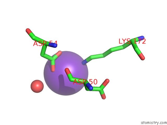
Mono view
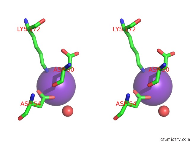
Stereo pair view
Mono view
Stereo pair view
A full contact list of Potassium with other atoms in the K binding
site number 1 of Crystal Structure of A Prolyl 4-Hydroxylase Complexed with Alpha- Ketoglutarate From the Pathogenic Bacterium Bacillus Anthracis in C2221 within 5.0Å range:
|
Potassium binding site 2 out of 6 in 5hv4
Go back to
Potassium binding site 2 out
of 6 in the Crystal Structure of A Prolyl 4-Hydroxylase Complexed with Alpha- Ketoglutarate From the Pathogenic Bacterium Bacillus Anthracis in C2221
Mono view

Stereo pair view
Mono view
Stereo pair view
A full contact list of Potassium with other atoms in the K binding
site number 2 of Crystal Structure of A Prolyl 4-Hydroxylase Complexed with Alpha- Ketoglutarate From the Pathogenic Bacterium Bacillus Anthracis in C2221 within 5.0Å range:
|
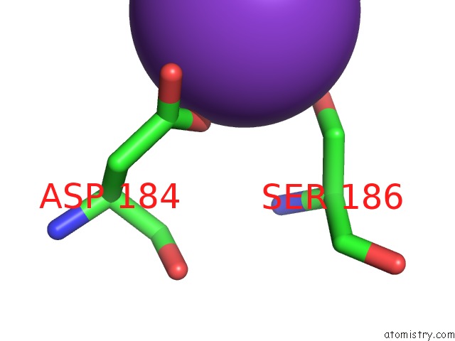
Potassium binding site 3 out of 6 in 5hv4
Go back to
Potassium binding site 3 out
of 6 in the Crystal Structure of A Prolyl 4-Hydroxylase Complexed with Alpha- Ketoglutarate From the Pathogenic Bacterium Bacillus Anthracis in C2221

Mono view
Stereo pair view
Mono view
Stereo pair view
A full contact list of Potassium with other atoms in the K binding
site number 3 of Crystal Structure of A Prolyl 4-Hydroxylase Complexed with Alpha- Ketoglutarate From the Pathogenic Bacterium Bacillus Anthracis in C2221 within 5.0Å range:
|
Potassium binding site 4 out of 6 in 5hv4
Go back to
Potassium binding site 4 out
of 6 in the Crystal Structure of A Prolyl 4-Hydroxylase Complexed with Alpha- Ketoglutarate From the Pathogenic Bacterium Bacillus Anthracis in C2221

Mono view

Stereo pair view
Mono view
Stereo pair view
A full contact list of Potassium with other atoms in the K binding
site number 4 of Crystal Structure of A Prolyl 4-Hydroxylase Complexed with Alpha- Ketoglutarate From the Pathogenic Bacterium Bacillus Anthracis in C2221 within 5.0Å range:
|
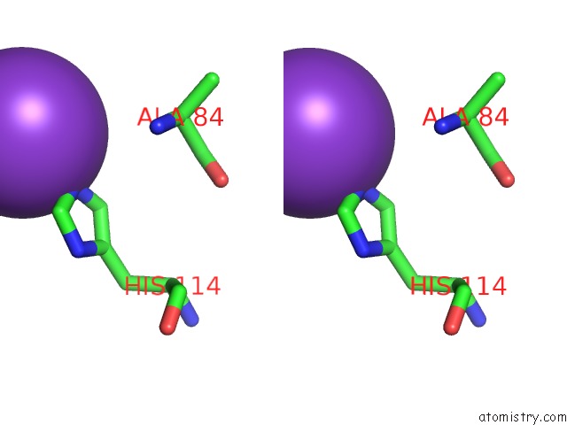
Potassium binding site 5 out of 6 in 5hv4
Go back to
Potassium binding site 5 out
of 6 in the Crystal Structure of A Prolyl 4-Hydroxylase Complexed with Alpha- Ketoglutarate From the Pathogenic Bacterium Bacillus Anthracis in C2221

Mono view

Stereo pair view
Mono view
Stereo pair view
A full contact list of Potassium with other atoms in the K binding
site number 5 of Crystal Structure of A Prolyl 4-Hydroxylase Complexed with Alpha- Ketoglutarate From the Pathogenic Bacterium Bacillus Anthracis in C2221 within 5.0Å range:
|
Potassium binding site 6 out of 6 in 5hv4
Go back to
Potassium binding site 6 out
of 6 in the Crystal Structure of A Prolyl 4-Hydroxylase Complexed with Alpha- Ketoglutarate From the Pathogenic Bacterium Bacillus Anthracis in C2221

Mono view

Stereo pair view
Mono view
Stereo pair view
A full contact list of Potassium with other atoms in the K binding
site number 6 of Crystal Structure of A Prolyl 4-Hydroxylase Complexed with Alpha- Ketoglutarate From the Pathogenic Bacterium Bacillus Anthracis in C2221 within 5.0Å range:
|
Reference:
N.J.Schnicker,
M.Dey.
Structural Analysis of Cofactor Binding For A Prolyl 4-Hydroxylase From the Pathogenic Bacterium Bacillus Anthracis. Acta Crystallogr D Struct V. 72 675 2016BIOL.
ISSN: ISSN 2059-7983
PubMed: 27139630
DOI: 10.1107/S2059798316004198
Page generated: Sat Aug 9 09:14:52 2025
ISSN: ISSN 2059-7983
PubMed: 27139630
DOI: 10.1107/S2059798316004198
Last articles
Na in 4CDUNa in 4CD5
Na in 4CDQ
Na in 4CCY
Na in 4CBK
Na in 4CD4
Na in 4CCS
Na in 4CBC
Na in 4CCG
Na in 4CBX