Potassium »
PDB 5cbv-5dea »
5dav »
Potassium in PDB 5dav: Fe(II)/(Alpha)Ketoglutarate-Dependent Dioxygenase Asqj in Complex with 4-Methoxydehydrocyclopeptin
Protein crystallography data
The structure of Fe(II)/(Alpha)Ketoglutarate-Dependent Dioxygenase Asqj in Complex with 4-Methoxydehydrocyclopeptin, PDB code: 5dav
was solved by
M.Groll,
A.Braeuer,
with X-Ray Crystallography technique. A brief refinement statistics is given in the table below:
| Resolution Low / High (Å) | 15.00 / 1.80 |
| Space group | C 2 2 21 |
| Cell size a, b, c (Å), α, β, γ (°) | 73.050, 120.770, 66.660, 90.00, 90.00, 90.00 |
| R / Rfree (%) | 16.4 / 19.6 |
Other elements in 5dav:
The structure of Fe(II)/(Alpha)Ketoglutarate-Dependent Dioxygenase Asqj in Complex with 4-Methoxydehydrocyclopeptin also contains other interesting chemical elements:
| Nickel | (Ni) | 1 atom |
| Bromine | (Br) | 2 atoms |
Potassium Binding Sites:
The binding sites of Potassium atom in the Fe(II)/(Alpha)Ketoglutarate-Dependent Dioxygenase Asqj in Complex with 4-Methoxydehydrocyclopeptin
(pdb code 5dav). This binding sites where shown within
5.0 Angstroms radius around Potassium atom.
In total only one binding site of Potassium was determined in the Fe(II)/(Alpha)Ketoglutarate-Dependent Dioxygenase Asqj in Complex with 4-Methoxydehydrocyclopeptin, PDB code: 5dav:
In total only one binding site of Potassium was determined in the Fe(II)/(Alpha)Ketoglutarate-Dependent Dioxygenase Asqj in Complex with 4-Methoxydehydrocyclopeptin, PDB code: 5dav:
Potassium binding site 1 out of 1 in 5dav
Go back to
Potassium binding site 1 out
of 1 in the Fe(II)/(Alpha)Ketoglutarate-Dependent Dioxygenase Asqj in Complex with 4-Methoxydehydrocyclopeptin
Mono view
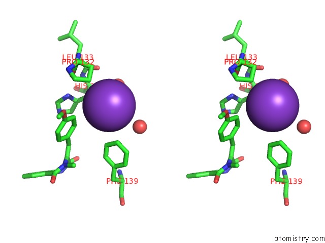
Stereo pair view
Mono view
Stereo pair view
A full contact list of Potassium with other atoms in the K binding
site number 1 of Fe(II)/(Alpha)Ketoglutarate-Dependent Dioxygenase Asqj in Complex with 4-Methoxydehydrocyclopeptin within 5.0Å range:
|
Reference:
A.Brauer,
P.Beck,
L.Hintermann,
M.Groll.
Structure of the Dioxygenase Asqj: Mechanistic Insights Into A One-Pot Multistep Quinolone Antibiotic Biosynthesis. Angew.Chem.Int.Ed.Engl. V. 55 422 2016.
ISSN: ESSN 1521-3773
PubMed: 26553478
DOI: 10.1002/ANIE.201507835
Page generated: Mon Aug 12 13:12:20 2024
ISSN: ESSN 1521-3773
PubMed: 26553478
DOI: 10.1002/ANIE.201507835
Last articles
K in 2VDDK in 2V5X
K in 2V5W
K in 2UXD
K in 2UYY
K in 2UUB
K in 2UU9
K in 2UXB
K in 2UUA
K in 2QYO