Potassium »
PDB 5cbv-5dea »
5cbv »
Potassium in PDB 5cbv: Human Cyclophilin D Complexed with Inhibitor
Protein crystallography data
The structure of Human Cyclophilin D Complexed with Inhibitor, PDB code: 5cbv
was solved by
R.P.Gibson,
E.Shore,
N.Kershaw,
M.Awais,
A.Javed,
D.Latawiec,
S.Pandalaneni,
L.Wen,
N.Berry,
P.O'neill,
R.Sutton,
L.Y.Lian,
with X-Ray Crystallography technique. A brief refinement statistics is given in the table below:
| Resolution Low / High (Å) | 38.20 / 1.80 |
| Space group | P 43 21 2 |
| Cell size a, b, c (Å), α, β, γ (°) | 57.430, 57.430, 113.040, 90.00, 90.00, 90.00 |
| R / Rfree (%) | 16.3 / 19.2 |
Potassium Binding Sites:
The binding sites of Potassium atom in the Human Cyclophilin D Complexed with Inhibitor
(pdb code 5cbv). This binding sites where shown within
5.0 Angstroms radius around Potassium atom.
In total only one binding site of Potassium was determined in the Human Cyclophilin D Complexed with Inhibitor, PDB code: 5cbv:
In total only one binding site of Potassium was determined in the Human Cyclophilin D Complexed with Inhibitor, PDB code: 5cbv:
Potassium binding site 1 out of 1 in 5cbv
Go back to
Potassium binding site 1 out
of 1 in the Human Cyclophilin D Complexed with Inhibitor
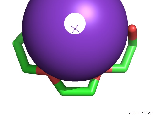
Mono view
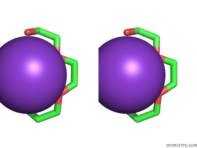
Stereo pair view
Mono view
Stereo pair view
A full contact list of Potassium with other atoms in the K binding
site number 1 of Human Cyclophilin D Complexed with Inhibitor within 5.0Å range:
|
Reference:
R.P.Gibson,
E.Shore,
N.Kershaw,
M.Awais,
A.Javed,
D.Latawiec,
S.Pandalaneni,
L.Wen,
N.Berry,
P.O'neill,
R.Sutton,
L.Y.Lian.
Human Cyclophilin D Complexed with Inhibitor To Be Published.
Page generated: Sat Aug 9 08:39:33 2025
Last articles
Mg in 6ZXSMg in 7A0Q
Mg in 7A0P
Mg in 7A0C
Mg in 721P
Mg in 6ZXH
Mg in 6ZXG
Mg in 6ZZ6
Mg in 6ZYM
Mg in 6ZY9