Potassium »
PDB 4xs6-4zum »
4zqr »
Potassium in PDB 4zqr: Crystal Structure of the Catalytic Domain of the Inosine Monophosphate Dehydrogenase From Mycobacterium Tuberculosis
Enzymatic activity of Crystal Structure of the Catalytic Domain of the Inosine Monophosphate Dehydrogenase From Mycobacterium Tuberculosis
All present enzymatic activity of Crystal Structure of the Catalytic Domain of the Inosine Monophosphate Dehydrogenase From Mycobacterium Tuberculosis:
1.1.1.205;
1.1.1.205;
Protein crystallography data
The structure of Crystal Structure of the Catalytic Domain of the Inosine Monophosphate Dehydrogenase From Mycobacterium Tuberculosis, PDB code: 4zqr
was solved by
Y.Kim,
M.Makowska-Grzyska,
M.Gu,
M.Kavitha,
L.Hedstrom,
W.F.Anderson,
A.Joachimiak,
Center For Structural Genomics Of Infectious Diseases(Csgid),
with X-Ray Crystallography technique. A brief refinement statistics is given in the table below:
| Resolution Low / High (Å) | 35.73 / 1.69 |
| Space group | P 1 |
| Cell size a, b, c (Å), α, β, γ (°) | 75.217, 75.233, 75.291, 108.30, 108.25, 111.88 |
| R / Rfree (%) | 15.1 / 18.3 |
Potassium Binding Sites:
The binding sites of Potassium atom in the Crystal Structure of the Catalytic Domain of the Inosine Monophosphate Dehydrogenase From Mycobacterium Tuberculosis
(pdb code 4zqr). This binding sites where shown within
5.0 Angstroms radius around Potassium atom.
In total 4 binding sites of Potassium where determined in the Crystal Structure of the Catalytic Domain of the Inosine Monophosphate Dehydrogenase From Mycobacterium Tuberculosis, PDB code: 4zqr:
Jump to Potassium binding site number: 1; 2; 3; 4;
In total 4 binding sites of Potassium where determined in the Crystal Structure of the Catalytic Domain of the Inosine Monophosphate Dehydrogenase From Mycobacterium Tuberculosis, PDB code: 4zqr:
Jump to Potassium binding site number: 1; 2; 3; 4;
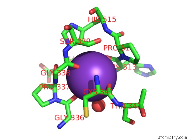
Potassium binding site 1 out of 4 in 4zqr
Go back to
Potassium binding site 1 out
of 4 in the Crystal Structure of the Catalytic Domain of the Inosine Monophosphate Dehydrogenase From Mycobacterium Tuberculosis

Mono view
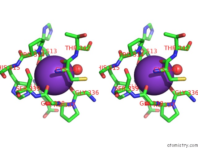
Stereo pair view
Mono view
Stereo pair view
A full contact list of Potassium with other atoms in the K binding
site number 1 of Crystal Structure of the Catalytic Domain of the Inosine Monophosphate Dehydrogenase From Mycobacterium Tuberculosis within 5.0Å range:
|
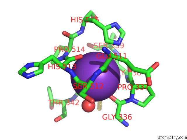
Potassium binding site 2 out of 4 in 4zqr
Go back to
Potassium binding site 2 out
of 4 in the Crystal Structure of the Catalytic Domain of the Inosine Monophosphate Dehydrogenase From Mycobacterium Tuberculosis
Mono view

Stereo pair view
Mono view
Stereo pair view
A full contact list of Potassium with other atoms in the K binding
site number 2 of Crystal Structure of the Catalytic Domain of the Inosine Monophosphate Dehydrogenase From Mycobacterium Tuberculosis within 5.0Å range:
|
Potassium binding site 3 out of 4 in 4zqr
Go back to
Potassium binding site 3 out
of 4 in the Crystal Structure of the Catalytic Domain of the Inosine Monophosphate Dehydrogenase From Mycobacterium Tuberculosis
Mono view

Stereo pair view
Mono view
Stereo pair view
A full contact list of Potassium with other atoms in the K binding
site number 3 of Crystal Structure of the Catalytic Domain of the Inosine Monophosphate Dehydrogenase From Mycobacterium Tuberculosis within 5.0Å range:
|
Potassium binding site 4 out of 4 in 4zqr
Go back to
Potassium binding site 4 out
of 4 in the Crystal Structure of the Catalytic Domain of the Inosine Monophosphate Dehydrogenase From Mycobacterium Tuberculosis
Mono view

Stereo pair view
Mono view
Stereo pair view
A full contact list of Potassium with other atoms in the K binding
site number 4 of Crystal Structure of the Catalytic Domain of the Inosine Monophosphate Dehydrogenase From Mycobacterium Tuberculosis within 5.0Å range:
|
Reference:
M.Makowska-Grzyska,
Y.Kim,
S.K.Gorla,
Y.Wei,
K.Mandapati,
M.Zhang,
N.Maltseva,
G.Modi,
H.I.Boshoff,
M.Gu,
C.Aldrich,
G.D.Cuny,
L.Hedstrom,
A.Joachimiak.
Mycobacterium Tuberculosis Impdh in Complexes with Substrates, Products and Antitubercular Compounds. Plos One V. 10 38976 2015.
ISSN: ESSN 1932-6203
PubMed: 26440283
DOI: 10.1371/JOURNAL.PONE.0138976
Page generated: Mon Aug 12 12:48:09 2024
ISSN: ESSN 1932-6203
PubMed: 26440283
DOI: 10.1371/JOURNAL.PONE.0138976
Last articles
Cu in 5D4JCu in 5DJT
Cu in 5D4H
Cu in 5D4I
Cu in 5CE9
Cu in 5CJ3
Cu in 5B7M
Cu in 5C92
Cu in 5C14
Cu in 5C0U