Potassium »
PDB 4qxg-4tog »
4rkj »
Potassium in PDB 4rkj: Crystal Structure of Thrombin Mutant S195T (Free Form)
Enzymatic activity of Crystal Structure of Thrombin Mutant S195T (Free Form)
All present enzymatic activity of Crystal Structure of Thrombin Mutant S195T (Free Form):
3.4.21.5;
3.4.21.5;
Protein crystallography data
The structure of Crystal Structure of Thrombin Mutant S195T (Free Form), PDB code: 4rkj
was solved by
A.L.Pelc,
Z.Chen,
D.W.Gohara,
A.D.Vogt,
N.Pozzi,
E.Di Cera,
with X-Ray Crystallography technique. A brief refinement statistics is given in the table below:
| Resolution Low / High (Å) | 40.00 / 1.70 |
| Space group | P 21 21 2 |
| Cell size a, b, c (Å), α, β, γ (°) | 61.415, 91.114, 50.553, 90.00, 90.00, 90.00 |
| R / Rfree (%) | 15.6 / 20.7 |
Potassium Binding Sites:
The binding sites of Potassium atom in the Crystal Structure of Thrombin Mutant S195T (Free Form)
(pdb code 4rkj). This binding sites where shown within
5.0 Angstroms radius around Potassium atom.
In total only one binding site of Potassium was determined in the Crystal Structure of Thrombin Mutant S195T (Free Form), PDB code: 4rkj:
In total only one binding site of Potassium was determined in the Crystal Structure of Thrombin Mutant S195T (Free Form), PDB code: 4rkj:
Potassium binding site 1 out of 1 in 4rkj
Go back to
Potassium binding site 1 out
of 1 in the Crystal Structure of Thrombin Mutant S195T (Free Form)
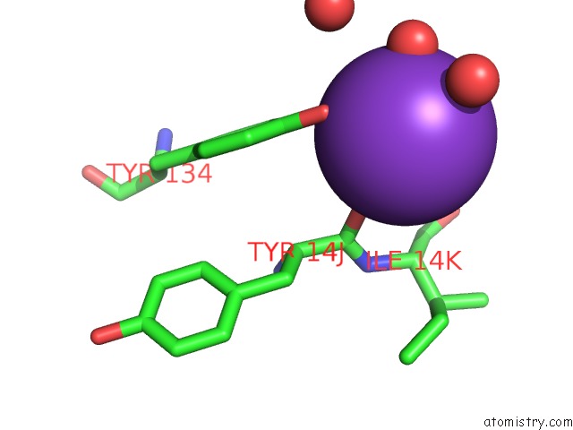
Mono view
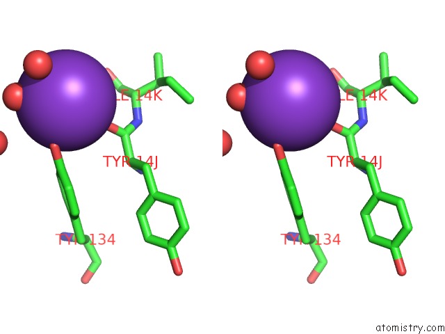
Stereo pair view
Mono view
Stereo pair view
A full contact list of Potassium with other atoms in the K binding
site number 1 of Crystal Structure of Thrombin Mutant S195T (Free Form) within 5.0Å range:
|
Reference:
L.A.Pelc,
Z.Chen,
D.W.Gohara,
A.D.Vogt,
N.Pozzi,
E.Di Cera.
Why Ser and Not Thr Brokers Catalysis in the Trypsin Fold. Biochemistry V. 54 1457 2015.
ISSN: ISSN 0006-2960
PubMed: 25664608
DOI: 10.1021/ACS.BIOCHEM.5B00014
Page generated: Sat Aug 9 07:48:19 2025
ISSN: ISSN 0006-2960
PubMed: 25664608
DOI: 10.1021/ACS.BIOCHEM.5B00014
Last articles
Na in 3MBBNa in 3MA9
Na in 3MAO
Na in 3MAT
Na in 3M7K
Na in 3M1H
Na in 3M9Y
Na in 3M8A
Na in 3M92
Na in 3M1A