Potassium »
PDB 4mlz-4pjo »
4nmc »
Potassium in PDB 4nmc: Crystal Structure of Oxidized Proline Utilization A (Puta) From Geobacter Sulfurreducens Pca Complexed with Zwittergent 3-12
Enzymatic activity of Crystal Structure of Oxidized Proline Utilization A (Puta) From Geobacter Sulfurreducens Pca Complexed with Zwittergent 3-12
All present enzymatic activity of Crystal Structure of Oxidized Proline Utilization A (Puta) From Geobacter Sulfurreducens Pca Complexed with Zwittergent 3-12:
1.2.1.88; 1.5.1.12; 1.5.99.8;
1.2.1.88; 1.5.1.12; 1.5.99.8;
Protein crystallography data
The structure of Crystal Structure of Oxidized Proline Utilization A (Puta) From Geobacter Sulfurreducens Pca Complexed with Zwittergent 3-12, PDB code: 4nmc
was solved by
H.Singh,
J.J.Tanner,
with X-Ray Crystallography technique. A brief refinement statistics is given in the table below:
| Resolution Low / High (Å) | 47.04 / 1.90 |
| Space group | P 21 21 21 |
| Cell size a, b, c (Å), α, β, γ (°) | 94.078, 157.207, 190.762, 90.00, 90.00, 90.00 |
| R / Rfree (%) | 18 / 21.3 |
Potassium Binding Sites:
The binding sites of Potassium atom in the Crystal Structure of Oxidized Proline Utilization A (Puta) From Geobacter Sulfurreducens Pca Complexed with Zwittergent 3-12
(pdb code 4nmc). This binding sites where shown within
5.0 Angstroms radius around Potassium atom.
In total 2 binding sites of Potassium where determined in the Crystal Structure of Oxidized Proline Utilization A (Puta) From Geobacter Sulfurreducens Pca Complexed with Zwittergent 3-12, PDB code: 4nmc:
Jump to Potassium binding site number: 1; 2;
In total 2 binding sites of Potassium where determined in the Crystal Structure of Oxidized Proline Utilization A (Puta) From Geobacter Sulfurreducens Pca Complexed with Zwittergent 3-12, PDB code: 4nmc:
Jump to Potassium binding site number: 1; 2;
Potassium binding site 1 out of 2 in 4nmc
Go back to
Potassium binding site 1 out
of 2 in the Crystal Structure of Oxidized Proline Utilization A (Puta) From Geobacter Sulfurreducens Pca Complexed with Zwittergent 3-12
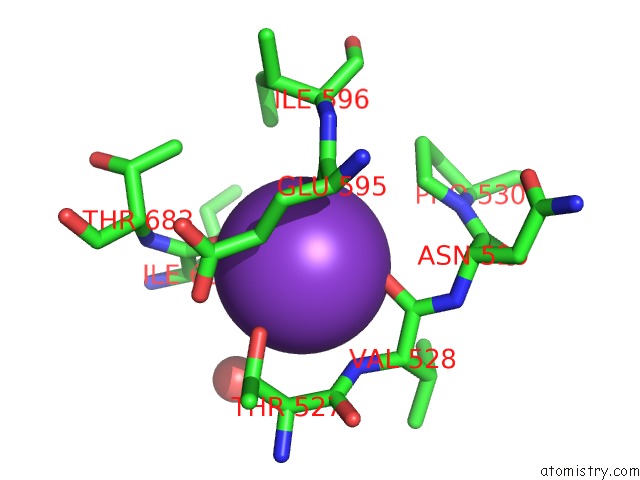
Mono view
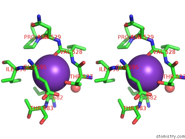
Stereo pair view
Mono view
Stereo pair view
A full contact list of Potassium with other atoms in the K binding
site number 1 of Crystal Structure of Oxidized Proline Utilization A (Puta) From Geobacter Sulfurreducens Pca Complexed with Zwittergent 3-12 within 5.0Å range:
|
Potassium binding site 2 out of 2 in 4nmc
Go back to
Potassium binding site 2 out
of 2 in the Crystal Structure of Oxidized Proline Utilization A (Puta) From Geobacter Sulfurreducens Pca Complexed with Zwittergent 3-12
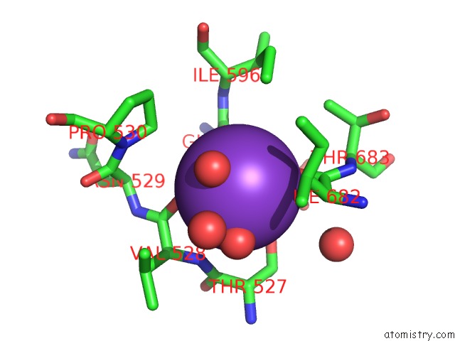
Mono view
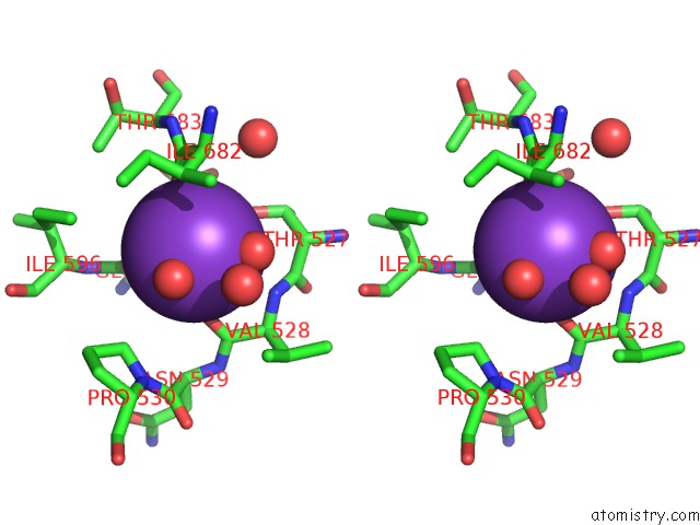
Stereo pair view
Mono view
Stereo pair view
A full contact list of Potassium with other atoms in the K binding
site number 2 of Crystal Structure of Oxidized Proline Utilization A (Puta) From Geobacter Sulfurreducens Pca Complexed with Zwittergent 3-12 within 5.0Å range:
|
Reference:
H.Singh,
B.W.Arentson,
D.F.Becker,
J.J.Tanner.
Structures of the Puta Peripheral Membrane Flavoenzyme Reveal A Dynamic Substrate-Channeling Tunnel and the Quinone-Binding Site. Proc.Natl.Acad.Sci.Usa V. 111 3389 2014.
ISSN: ISSN 0027-8424
PubMed: 24550478
DOI: 10.1073/PNAS.1321621111
Page generated: Sat Aug 9 07:32:03 2025
ISSN: ISSN 0027-8424
PubMed: 24550478
DOI: 10.1073/PNAS.1321621111
Last articles
Mo in 8BQTMo in 8CCQ
Mo in 8BTS
Mo in 8BQR
Mo in 8BQQ
Mo in 8BQP
Mo in 7Z5J
Mo in 7ZUB
Mo in 7Z0T
Mo in 7WY3