Potassium »
PDB 4l4d-4mkk »
4lz1 »
Potassium in PDB 4lz1: X-Ray Structure of the Complex Between Human Thrombin and the Tba Deletion Mutant Lacking Thymine 12 Nucleobase
Enzymatic activity of X-Ray Structure of the Complex Between Human Thrombin and the Tba Deletion Mutant Lacking Thymine 12 Nucleobase
All present enzymatic activity of X-Ray Structure of the Complex Between Human Thrombin and the Tba Deletion Mutant Lacking Thymine 12 Nucleobase:
3.4.21.5;
3.4.21.5;
Protein crystallography data
The structure of X-Ray Structure of the Complex Between Human Thrombin and the Tba Deletion Mutant Lacking Thymine 12 Nucleobase, PDB code: 4lz1
was solved by
A.Pica,
I.Russo Krauss,
A.Merlino,
F.Sica,
with X-Ray Crystallography technique. A brief refinement statistics is given in the table below:
| Resolution Low / High (Å) | 25.20 / 1.65 |
| Space group | P 1 |
| Cell size a, b, c (Å), α, β, γ (°) | 42.892, 44.484, 52.643, 65.24, 82.53, 63.72 |
| R / Rfree (%) | 15.5 / 19 |
Other elements in 4lz1:
The structure of X-Ray Structure of the Complex Between Human Thrombin and the Tba Deletion Mutant Lacking Thymine 12 Nucleobase also contains other interesting chemical elements:
| Sodium | (Na) | 1 atom |
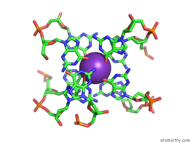
Potassium Binding Sites:
The binding sites of Potassium atom in the X-Ray Structure of the Complex Between Human Thrombin and the Tba Deletion Mutant Lacking Thymine 12 Nucleobase
(pdb code 4lz1). This binding sites where shown within
5.0 Angstroms radius around Potassium atom.
In total only one binding site of Potassium was determined in the X-Ray Structure of the Complex Between Human Thrombin and the Tba Deletion Mutant Lacking Thymine 12 Nucleobase, PDB code: 4lz1:
In total only one binding site of Potassium was determined in the X-Ray Structure of the Complex Between Human Thrombin and the Tba Deletion Mutant Lacking Thymine 12 Nucleobase, PDB code: 4lz1:
Potassium binding site 1 out of 1 in 4lz1
Go back to
Potassium binding site 1 out
of 1 in the X-Ray Structure of the Complex Between Human Thrombin and the Tba Deletion Mutant Lacking Thymine 12 Nucleobase
Mono view

Stereo pair view
Mono view
Stereo pair view
A full contact list of Potassium with other atoms in the K binding
site number 1 of X-Ray Structure of the Complex Between Human Thrombin and the Tba Deletion Mutant Lacking Thymine 12 Nucleobase within 5.0Å range:
|
Reference:
A.Pica,
I.Russo Krauss,
A.Merlino,
S.Nagatoishi,
N.Sugimoto,
F.Sica.
Dissecting the Contribution of Thrombin Exosite I in the Recognition of Thrombin Binding Aptamer. Febs J. V. 280 6581 2013.
ISSN: ISSN 1742-464X
PubMed: 24128303
DOI: 10.1111/FEBS.12561
Page generated: Sat Aug 9 07:25:56 2025
ISSN: ISSN 1742-464X
PubMed: 24128303
DOI: 10.1111/FEBS.12561
Last articles
Na in 7RQVNa in 7RQX
Na in 7RQU
Na in 7RPH
Na in 7RPK
Na in 7RPI
Na in 7ROF
Na in 7ROO
Na in 7RNQ
Na in 7RNP