Potassium »
PDB 4gx1-4ije »
4h6u »
Potassium in PDB 4h6u: Tubulin Acetyltransferase Mutant
Enzymatic activity of Tubulin Acetyltransferase Mutant
All present enzymatic activity of Tubulin Acetyltransferase Mutant:
2.3.1.108;
2.3.1.108;
Protein crystallography data
The structure of Tubulin Acetyltransferase Mutant, PDB code: 4h6u
was solved by
A.Roll-Mecak,
V.Kizub,
A.Szyk,
with X-Ray Crystallography technique. A brief refinement statistics is given in the table below:
| Resolution Low / High (Å) | 29.06 / 2.45 |
| Space group | P 43 21 2 |
| Cell size a, b, c (Å), α, β, γ (°) | 58.128, 58.128, 225.070, 90.00, 90.00, 90.00 |
| R / Rfree (%) | 19.6 / 24.8 |
Potassium Binding Sites:
The binding sites of Potassium atom in the Tubulin Acetyltransferase Mutant
(pdb code 4h6u). This binding sites where shown within
5.0 Angstroms radius around Potassium atom.
In total only one binding site of Potassium was determined in the Tubulin Acetyltransferase Mutant, PDB code: 4h6u:
In total only one binding site of Potassium was determined in the Tubulin Acetyltransferase Mutant, PDB code: 4h6u:
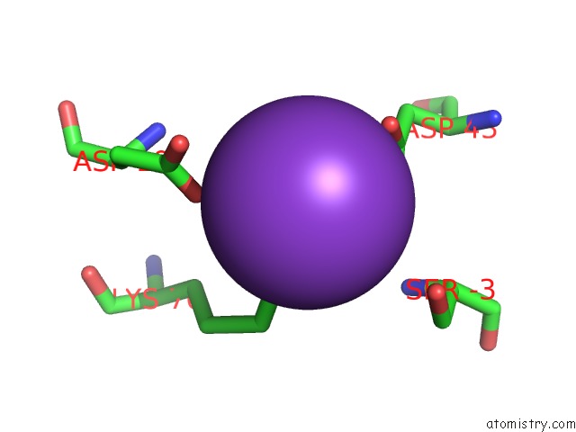
Potassium binding site 1 out of 1 in 4h6u
Go back to
Potassium binding site 1 out
of 1 in the Tubulin Acetyltransferase Mutant
Mono view

Stereo pair view
Mono view
Stereo pair view
A full contact list of Potassium with other atoms in the K binding
site number 1 of Tubulin Acetyltransferase Mutant within 5.0Å range:
|
Reference:
V.Kormendi,
A.Szyk,
G.Piszczek,
A.Roll-Mecak.
Crystal Structures of Tubulin Acetyltransferase Reveal A Conserved Catalytic Core and the Plasticity of the Essential N Terminus. J.Biol.Chem. V. 287 41569 2012.
ISSN: ISSN 0021-9258
PubMed: 23105108
DOI: 10.1074/JBC.C112.421222
Page generated: Sat Aug 9 06:56:33 2025
ISSN: ISSN 0021-9258
PubMed: 23105108
DOI: 10.1074/JBC.C112.421222
Last articles
Mn in 7BHIMn in 7BDW
Mn in 7BBM
Mn in 7B4S
Mn in 7B4R
Mn in 7B0H
Mn in 7AVW
Mn in 7B1S
Mn in 7AZR
Mn in 7AUA